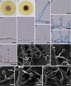
Phaeoacremonium italicum A. Carlucci & M.L. Raimondo., in Raimondo et al., Mycologia 106(6), 1123 (2014), Index Fungorum Number: 808660
Associated with stem wilt disease of T. grandis. Sexual morph: Undetermined. Asexual morph: Structures on MEA: Mycelium 1–2.6μm broad, branched, septate, single or in bundles, partly superficial, partly immersed, hyaline to pale brown, verruculose. Conidiophores up to 40μm long, 1– 2.8 diam, erect to slightly curved, up to 2–3-septate, swollen at the base, tapering at the apex, occasionally bearing 2 lateral phialides next to the terminal phialide, wide, hyaline to pale brown, each newly proliferated segment swollen at the base. Phialides terminal or lateral, mostly monophialidic, occasionally polyphialidic; type I phialides (14–)16–19(−26) high × (2.2–)2.3–2.4(−2.7) μm (x = 18×2.4μm) , adelophialide, subcylindrical, no basal septum; type II phialides (14–) 20–24 (−31) high × (1.9–)2.3 2.5(−2.9) μm (x = 20×2.4μm), predominant, elongate-ampulliform attenuated at the base to subcylindrical, constricted at the base, tapering towards the apex; type III phialides (13–)22–29(−41) high× (2–)2.5–2.7(−3.1)μm (x = 25×2.6μm), subcylindrical to navicular. Conidia (1–)3.6–3.9(−4.7)× (1–)1.5–1.7(−2) μm (x = 3.6 × 1.6 μm, n = 30), allantoid to oblong ellipsoidal, smooth to verrucose, hyaline.
Habitat: Known to inhabit Vitis vinifera (Raimondo et al. 2014) and associated with stem wilt disease of T. grandis (Doilom et al. 2017).
Known distribution: Italy (Raimondo et al. 2014) and Thailand (Doilom et al. 2017).
Material examined: THAILAND, Chiang Rai Province, Muang District, associated with stem wilt of T. grandis, 25 January 2013, M. Doilom & E.Wongsakul, MFLU 15–3427, dry culture, living culture MFLUCC 13–0336, MKT 104/1, GenBank Accession No: Actin: KU194224, TUB: KU194225.
Notes: It would appear that it is unlikely that Phaeoacremonium italicum (MFLUCC 13–0336) is the same as Pm. italicum as reported in Raimondo et al. (2014) due to differences in host and geography. In addition, it did not produce a yellow pigment on MEA and PDA. Differences are evident also in the conidiophores of Pm. italicum (MFLUCC 13–0336) which are unbranched to branched, but unbranched for ex-type culture (Raimondo et al. 2014). However, the type II phialides in the collections in the present study predominate and the isolate MFLUCC 13–0336 is closely related to Pm. italicum based on DNA sequence data.
FIG: Phaeoacremonium italicum (MFLU 15–3427). a, b Colony on MEA after 16 days (a = above view, b = below view). c, e, f Conidiophores. d Type I phialides. h Type II phialides with conidia. g Type III phialides. j Type III phialides with conidia. k–o Conidiophores with conidia. Notes: k–o Micrograph from Scanning Electron Microscope (SEM). c, h, i Stained in lactophenol cotton blue. Scale bars: c, d, h, j–l, n, o = 10μm, e–g, i, m = 5μm

