Sanguinoderma rude (Berk.) Y.F. Sun, D.H. Costa & B.K. Cui, comb. nov.
MycoBank number: MB 828446; Index Fungorum number: IF 828446; Facesoffungi number: FoF; Fig. 8o – p, 16
Basionym. Polyporus rudis Berk., Ann. Mag. Nat. Hist. 3: 323. 1839.
≡ Amauroderma rude (Berk.) Torrend, Brotéria, Sér. Bot. 18: 127. 1920.
Basidiomata annual, lateral stipitate to almost sessile, corky. Pileus single, suborbicular to flabelliform, up to 7.5 cm diam and 1.7 cm thick. Pileal surface dark brown to ferruginous, dull, glabrous, with faintly concentric zones and radial wrinkles; mar- gin acute to obtuse, entire, wavy and incurved when dry. Pore surface pale grey to greyish yellow, colour changing to blood red when bruised, then quickly darkening; pores subcircular to irregular, 1– 4 per mm; dissepiments slightly thick, entire. Context woody colour to pale brown, without resinous lines, soft corky, up to 9 mm thick. Tubes darker than pore surface, hard corky, up to 8 mm long. Stipe concolorous with pileal surface, cylindrical and hollow, up to 5 cm long and 4 mm diam. Hyphal system trimitic; generative hyphae with clamp connections, all hyphae IKI–, CB+; tissues darkening in KOH. Generative hyphae in context colourless, thin- to slightly thick-walled, 2 – 4 μm diam; skeletal hyphae in context pale yellow, thick-walled with
a wide to narrow lumen or subsolid, arboriform branched and flexuous, 2 – 8 μm diam; binding hyphae in context pale yellow, subsolid, branched and flexuous, 1– 2 μm diam. Generative hyphae in tubes colourless, thin- to slightly thick-walled, 2 – 4 μm diam; skeletal hyphae in tubes pale yellow, thick-walled with a wide to narrow lumen or subsolid, arboriform branched and flexuous, 3 – 6 μm diam; binding hyphae in tubes pale yellow, subsolid, 1– 2 μm diam. Pileal cover composed of clamped generative hyphae, thin- to thick-walled, apical cells clavate, faintly inflated, flexuous, yellowish brown, about 40 – 80 × 4–6 μm, forming an irregular palisade. Cystidia or cystidioles absent. Basidia barrel-shaped to clavate, colourless, thin-walled, 13– 33 × 12 – 28 μm; basidioles suborbicular to clavate, colourless, thin-walled, 15 – 25 × 8 –19 μm. Basidiospores subglobose to broadly ellipsoid, pale yellow, IKI–, CB+, with double and medially thick walls, exospore wall smooth, endospore wall with conspicuous spinules, (9 –)9.5 –12.5(–12.9) × 8 –10.7(–11) μm, L = 11.18 μm, W = 9.5 μm, Q = 1.17–1.19 (n = 60/2). Under SEM, exospore wall alveolate to semi-reticulate with small and deep pits, endospore wall with short and slightly thick columnar spinules tightly arranged.
Specimens examined. AUSTRALIA, Victoria State, Eastern Highlands, on rot- ten stump, 20 Mar. 1994, N.H. Sinnott, MEL 2028873 (MEL); East Gippsland, on buried wood, 27 Mar. 2002, K.R. Thiele, MEL 2150776 (MEL); Eastern Highlands, on stump of Eucalyptus, 12 May 2003, S.H. Lewis, MEL 2231602 (MEL); Gippsland Plain, on rotten stump of shrub, 4 May 2007, H. Strand, MEL 2317411 (MEL); Fairfield, on rotten stump of Acacia, 23 Apr. 2009, N.H. Sinnott, MEL 2335651 (MEL); Valley Reserve, on rotten wood, 14 July 2012, N.G. Karunajeewa, MEL 2362204 (MEL); Tasmania, on base of dead Eucalyptus, 12 May 2018, B.K. Cui, Cui 16592 (BJFC).
Notes — Amauroderma rude was described from Tasmania as Polyporus rudis (Berkeley 1839). We have examined the specimens collected from the type locality of Tasmania and mainland in Australia. It can be characterized by its big pores (1– 4 per mm) with slightly thick and entire dissepiments, and alveolate to semi-reticulate exospore wall with small and deep pits (Fig. 8o – p). Amauroderma rude was shown as a distinct well-supported lineage in Sanguinoderma (Fig. 1, 2). San guinoderma rude is similar to S. perplexum by the subglobose to broadly ellipsoid basidiospores, but S. perplexum differs from S. rude by the woody hard basidiomata, small pores (5 – 6 per mm) with medially thick dissepiments and vermiculate to semi-reticulate exospore wall with long and thick endospore spinules (Fig. 8k – l). Amauroderma intermedium is quite similar to S. rude both in macro- and micro-morphology (Gomes-Silva et al. 2015), but it can be distinguished by its Neotropical distribution, darker context and colour-unchanging pore surface when bruised (Torrend 1920, Ryvarden 2004).
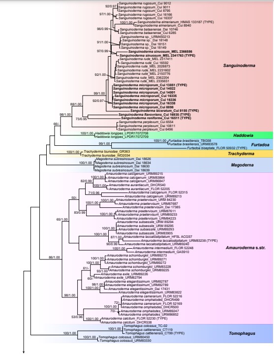
Fig. 1 ML analyses of Ganodermataceae based on dataset of ITS+nLSU+RPB1+TEF. ML bootstrap values higher than 50 % and Bayesian posterior probabilities values more than 0.95 are shown. New species are in bold.
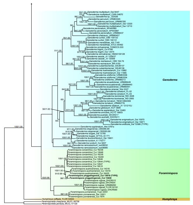
Fig. 1 (cont.)
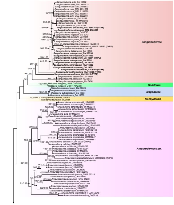
Fig. 2 ML analyses of Ganodermataceae based on dataset of ITS+nLSU+RPB1+RPB2+TEF+TUB. ML bootstrap values higher than 50 % and Bayesian posterior probabilities values more than 0.95 are shown. New species are in bold.
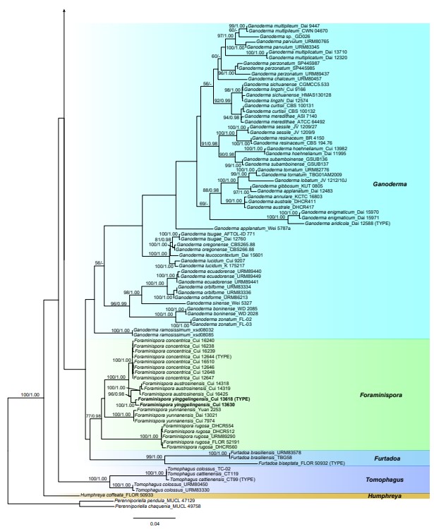
Fig. 2 (cont.)

Fig. 8 Scanning Electron Micrograph (SEM) of basidiospores of Sanguinoderma. a– b. S. bataanense (Dai 10746); c– d. S. elmerianum (HMAS 133187); e– f. S. flavovirens (Cui 16935); g– h. S. laceratum (Cui 8155); i– j. S. microporum (Cui 13851); k– l. S. perplexum (Dai 10811); m– n. S. reniforme (Cui 16511); o– p. S. rude (MEL 2150776); q– r. S. rugosum (Cui 16377); s– t. S. sinuosum (MEL 2366586). a, c, e, g, i, k, m, o, q, s: General view showing exospore wall; b, d, f, h, j, l, n, p, r, t: detail in endospore ornamentation. — Scale bars: a– t = 2 μm.
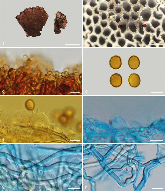
Fig. 16 Basidiomata and microscopic structures of Sanguinoderma rude (MEL 2335651). a. Basidiomata; b. pores; c. apical cells from pileal cover; d. basidio- spores; e. basidia; f. basidioles; g. skeletal hyphae from tubes; h. skeletal hyphae from context. — Scale bars: a = 3 cm; b = 0.5 mm; c– h = 10 µm.
