Pseudostomiopeltis xishuangbannaensis Phookamsak, Hongsanan, Wanas. & Bhat, in Hongsanan, Phookamsak, Bhat, Wanasinghe, Promputtha, Suwannarach, Sandamali, Lumyong, Xu & Xie, Frontiers in Cellular and Infection Microbiology 13(no. 1252387): 9 (2023)
Index Fungorum number: IF 900640; MycoBank number: MB 900640; Facesoffungi number: FoF 15866
Etymology – The specific epithet “xishuangbannaensis” refers to the locality, Xishuangbanna Dai Autonomous Prefecture, Yunnan, China, where the holotype was collected.
Holotype – KUN-HKAS 129044
Epiphytic or saprobic on dead leaves of an unidentified dicot, hypophyllous, visible as small, circular, black dots, easily removed from the host surface. Sexual morph: Undetermined. Asexual morph: Conidiomata 85–120 µm high, 70–135 µm, thyriothecial, scattered, or in a small group, hemispherical, dimidiate-scutate, uni-loculate, with a central, pore-like ostiole. Upper wall composed of a thin layer, of brown, radiating cells, with loosely irregular lobed cells at the margin. Peridium 1.5–4 µm wide, composed of 1–2 strata of textura angularis. Conidiophores reduced to the conidiogenous cells. Conidiogenous cells 2.5–5 × 1.5–3 µm (𝑥̄ = 3.9 × 2.3, n = 30), arising from the innermost wall cells of the conidiomata, hyaline, enteroblastic, phialidic, lageniform to ampulliform, determinate, discrete, smooth-walled, with minute channel and collarette. Conidia 7–9 × 2–4 µm (𝑥̄ = 8.1 × 3.2, n = 30), solitary, hyaline, ellipsoidal to oblong, slightly truncate at the base, with obtuse apex, aseptate, smooth-walled, occasionally with attached conidiogenous cell.
Culture characteristics – Conidia germinated on PDA within 24 h. Colonies on OA reaching 25–28 mm in diam. after two weeks at room temperature (15°C–20°C). Colonies dense, circular, flattened to slightly raised, surface smooth with an entire edge, fairly fluffy to floccose; from above gray at the margin, becoming dark gray toward the center, with cream conidial masses; from below dark gray to black at the margin, white-gray at the middle, dark gray to black at the center, radiating. Sporulation in OA after two weeks, forming dense, brown, compact hyphae on OA medium, with cream spore masses. Mycelium 1.5–3 µm thick, brown, branched, septate, with compact hyphae. Conidiophores 10–30 × 1–3 µm (𝑥̄ = 13.9 × 2.2, n = 30), hyaline, cylindrical to subcylindrical, septate, branched or unbranched, smooth-walled. Conidiogenous cells (5–)7–9 × 1–3 µm (𝑥̄ = 7.8 × 1.9, n = 30), hyaline, enteroblastic, phialidic, terminal, cylindrical to subcylindrical, aseptate, smooth-walled, with minute channel and collarette. Conidia 2.5–4 × 1–2 µm (𝑥̄ = 3 × 1.7, n = 30), solitary, hyaline, subglobose to ellipsoidal to oblong, with obtuse ends, aseptate, smooth-walled.
Material examined – China, Yunnan Province, Xishuangbanna Dai Autonomous Prefecture, Mengla County, Menglun, Xishuangbanna Tropical Botanical Garden, on dead leaves of an unidentified dicot, 11 January 2021, D.N. Wanasinghe, Xh5 (KUN-HKAS 129044, holotype), ex-type living culture, RPC 21-031 = KUNCC.
Notes – The NCBI nucleotide BLAST search of ITS sequence indicated that Pseudostomiopeltis xishuangbannaensis (RPC 21-031) is similar to Stomiopeltis sp. T49A1c with 99.82% similarity (identities = 557/558 with no gap), Stomiopeltis sp. T36A1b with 97.32% similarity (identities = 545/560 with 5 gaps), Stomiopeltis sp. RS7 with 96.25% similarity (identities = 539/560 with 5 gaps), and S. phyllanthi MFLU 18-2115 with 92.36% similarity (identities = 447/484 with 9 gaps). Similarly, the NCBI nucleotide BLAST search of LSU sequence indicated that Ps. xishuangbannaensis (RPC 21-031) is similar to Cf. Stomiopeltis sp. RS7.2 with 100% similarity (identities = 765/765, with no gap), Cf. Stomiopeltis sp. RS7.1 with 99.87% similarity (identities = 764/765, with no gap), and Stomiopeltis sp. RS7 with 99.76% similarity (identities = 834/836, with 1 gap), while the LSU of S. phyllanthi MFLU 18-2115 is misidentified. The closest hit using the LSU sequence of S. phyllanthi is Meyerozyma guilliermondii culture CBS:8105 with 100% similarity. The phylogenetic analyses of a concatenated ITS and LSU sequence dataset revealed that Pseudostomiopeltis xishuangbannaensis (RPC 21-031) has a close relationship with Ps. phyllanthi MFLU 18-2115 (≡ Stomiopeltis phyllanthi). A nucleotide pairwise comparison of ITS sequence indicated that Ps. xishuangbannaensis (RPC 21-031) differs from Ps. phyllanthi (MFLU 18-2115) in 38/485 bp (7.8%). Morphologically, Ps. xishuangbannaensis (RPC 21-031) could not be compared with Ps. phyllanthi as they formed different morphs.
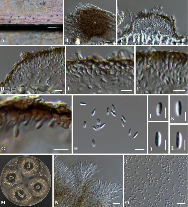
Figure 1 – Pseudostomiopeltis xishuangbannaensis (KUN-HKAS 129044, holotype). (A) The appearance of conidiomata on host substrate. (B) Upper view of conidioma. (C) Vertical section of conidioma. (D) Section through the peridium. (E–G) Conidiogenous cells bearing conidia, arising from the inner cavities (note: G = stained with Congo red). (H–L) Conidia. (M) Sporulation on OA with cream conidial masses. (N) Conidiophores and conidiogenous cells in vitro. (O) Conidia sporulated in vitro. Scale bars: (B, C) = 20 μm, (D–L, N, O) = 5 μm.