Roussoella siamensis Phookamsak, J.K. Liu & K.D. Hyde., in Liu et al. Phytotaxa 181(1): 1–33 (2014). Index Fungorum number: IF550665
Saprobic on dead bamboo culms. Sexual morph: Ascostromata 120–150 μm high, 620–750 μm diam., gregarious, immersed within cortex, visible as raised, brown to dark brown areas on the host surface, shield-shaped to continuously low convex, uni- to multi-loculate, coriaceous. Locules 70–120 μm high, 130–350 μm diam., scattered to clustered, gregarious, immersed in ascostromata, lenticular to subglobose, with a flattened base, glabrous, ostiole central, with a minute ostiole embedded beneath a clypeus, becoming pore-like opening on host surface at maturity. Peridium 7–15 μm wide, thinwalled of unequal thickness, thinner at the base, composed of several layers of flattened, brown to dark brown, compressed textura angularis pseudoparenchymatous cells. Hamathecium composed of dense, 1–2 μm wide, cellular pseudoparaphyses, indistinctly septate, not constricted at the septum, anastomosing and branching at the apex, embedded in a gelatinous matrix. Asci (67–)70–90(−94) × 6– 8 μm (x = 77.8 × 7.3 μm, n = 25), 8-spored, bitunicate, cylindrical, short to long pedicellate with furcate or footlike pedicel, apically rounded, with an indistinct ocular chamber. Ascospores (8.5–)10–12(−14) × 4–5 μm (x = 10.8 × 4.6 μm, n = 30), overlapping, uni-seriate, ellipsoidal to fusiform, with rounded ends, brown to dark brown, 1- septate, constricted at the septum, rough-walled, longitudinally ribbed. Asexual morph: Coelomycetous, produced on bamboo pieces on WA after 12 weeks. Conidiomata 360–940 μm high, 560–1300 μm diam., solitary to gregarious, clustered, semi-immersed to superficial, visible as black irregular pycnostroma, covered by hyphae on bamboo pieces, globose to subglobose, uni-loculate, becoming glabrous at maturity, indistinctly ostiolate. Conidiomata wall 20–45 μm wide, thin-walled, of equal thickness, composed of four strata, an inner layer comprising conidiogenous cells arising from a thin layer of hyaline cells, a stratum of 3–5 layers of dark brown to black, pseudoparenchymatous cells, arranged in a textura angularis, a median stratum comprising several layers of flattened, hyaline cells, arranged in textura angularis to textura prismatica, and an outer stratum comprising several layers of dark brown to black stromatic cells, arranged in textura intricata. Conidiophores arising from the basal cavity around conidiomata, unbranched, 0–1 septate, or reduced to conidiogenous cells. Conidiogenous cells 3– 6 (−10) × 2.5–5 μm (x = 5.3 × 3.7 μm, n = 30), enteroblastic, phialidic or broadly annellidic, determinate, discrete, unbranched, ampulliform to lageniform, or irregular in shape, hyaline, aseptate, smooth-walled, with distinct periclinal thickening. Conidia 4–5×3–4 μm (x = 4.8 × 3.4 μm, n = 50), globose to subglobose, initially hyaline, becoming brown, aseptate, rough-walled, verrucose.
Culture characters: Colonies on PDA fast growing, 55– 60 mm diam. after 4 weeks at 25–30 °C, colonies irregular, medium dense, slightly raised to umbonate, dull with undulate edge, floccose to woolly, slightly radiating, pale yellowish to brownish orange at the margin, white to cream medianly, pale yellowish at the centre from above, pale yellowish at the margin, brown to golden brown medianly, dark brown to black at the centre from below, produced yellowish brown pigment in agar.
Material examined: THAILAND, Chiang Rai Province, Muang District, Mae Fah Luang University campus grounds, on dead culms of Bambusa sp., 13 August 2010, R. Phookamsak, RP0065 (MFLU 11–0185, holotype), ex-type living cultures, MFLUCC 11–0149, KUMCC.
Notes: Roussoella siamensis was introduced by Liu et al. (2014), and lacked the asexual morph description. In this study, the ex-type culture produced a coeloemycetous asexual morph on bamboo pieces on water agar. Thus, we redescribe and illustrate this species based on morphological characters of both sexual and asexual morphs. The species was described as forming multi-loculate ascomata with cylindrical, bitunicate asci and brown, striate, didymosporous ascospores (Liu et al. 2014). Based on the morphological characters, Roussoella siamensis is similar to R. chiangraina and R. neopustulans (Liu et al. 2014). However, R. siamensis differs from R. chiangraina and R. neopustulans by forming multi-loculate ascostromata, and having larger asci, ascospores and conidia than R. chiangraina and R. neopustulans. Based on multi-gene phylogenetic analyses, R. siamensis forms a single basal clade (Fig. 19).
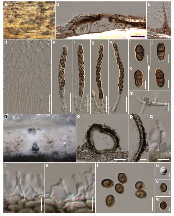
FIG Roussoella siamensis (MFLU 11–0185, holotype). a Appearance of ascostromata on host surface. b Vertical section through ascostroma. c Section through peridium between locules. d Pseudoparaphyses. e–h Asci. i–l Ascospores. m Germinating ascospore. n Conidiomata on bamboo pieces on WA. o Vertical section through conidioma. p Section through pycnidial wall. q–s Conidiogenous cells. t–w Conidia. Scale bars: b, o = 100 μm, c–h, p = 20 μm, m = 10 μm, i–l, q–s, t = 5 μm, u–w=2 μm