Diaporthe neoraonikayaporum Doilom, A.J. Dissanayake & K.D. Hyde, sp. nov., Index Fungorum number: IF551993
Etymology: In reference to its similarity to raonikayaporum.
Holotype: MFLU 15–3539 (dry culture)
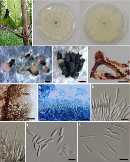
Associated with branch and twig dieback lesions on T. grandis. Sexual morph: Undetermined. Asexual morph: on PDA, C o n i d i o m a t a ( 690– ) 950–104 5 ( −119 0 ) μm × (805–)995–1110(−1285) μm (x = 925 × 1015 μm, n = 15), pycnidial, scattered or aggregated, black to reddish brown, subglobose or variable in shape, with an elongated black neck, with conidial mass; initially hyaline to yellowish, becoming white to cream or pink conidial droplets exuding from central ostioles; pycnidial wall composed of a textura angularis, comprising of two cell layers, outer layer; black to brown, thick-walled, inner layer; hyaline, thin-walled. Conidiophores 15–23×1.3–2μm, hyaline, branched to unbranched, cylindrical, septate, 1–3-septate, smooth, dense, straight or slightly curved, tapering towards the apex, wider at base. Conidiogenous cells 2.5–5.3×1.4–1.9μm, phialidic, cylindrical, terminal, with slight
tapering towards apex, 0.5–1μm diam. Alpha conidia (4–)4.9– 5.3(−6) μm×2–3μm (x = 5×2.4μm, n=30), with bi-guttulate to multi-guttulate, hyaline, ellipsoid, apex bluntly rounded, base obtuse to subtruncate, smooth. Beta conidia (13–)16–18(−21) μm×(1–)1.5–1.6(−1.8) μm (x = 16×1.5μm, n=30), filiform, hyaline, flexible to slightly curved, aseptate, base subtruncate. Gamma conidia (7–)8.5–9(−10.6)×(1.7–)2.2–2.3(−2.6) μm (x = 8.5×2.2μm, n=30), aseptate, hyaline, smooth, fusoid, with bi-guttulate to multi-guttulate, apex acutely rounded, base
subtruncate.
Culture characteristics: Pure culture was isolated by subbing hyphal tips growing from surface sterilized diseased material. Colonies on PDA reaching 64–77 mm diam. after 4 days in the dark at 25 °C, (x = 71, n= 5), lobate, medium dense, cottony, flat or effuse to low convex, with concentric rings, irregular margin, after 10 days white (4A1) from above and below, reaching the edge of the Petri-dish after 6–11 days. Habitat: Associated with branch and twig dieback lesions of T. grandis (current study). Known distribution: Thailand (current study).
Habitat: Associated with branch and twig dieback lesions of T. grandis (Doilom et al. 2017).
Known distribution: Thailand (Doilom et al. 2017).
Material examined: THAILAND, Chiang Rai Province, Mae Suai District, Mae Lao garden, on dieback lesion of T. grandis branches, 5 July 2014, (MFLU 15–3539, holotype (dry–culture)), living culture MFLUCC 14–1136, MKT 168/ 1, ICMP 21176, GenBank Accession No: CAL: KU749356, ITS: KU712449, TEF1: KU749369, TUB: KU743988; ibid with MFLUCC 14–1136, living culture MFLUCC 14–1137, MKT 168/7, GenBank Accession No: CAL: KU749357, ITS: KU712450, TEF1: KU749370, TUB: KU743989; Chiang Rai Province, Muang District, Doi Lan Subdistrict, on dieback lesion of T. grandis twigs, 14 June 2014, M. Doilom, (MFLU 15–3538, paratype (dry–culture)), ex-paratype living culture MFLUCC 14–1133, MKT 164/1, Accession No: CAL: KU749355, ITS: KU712448, TEF1: KU749368, TUB: KU743987.
Notes: Despite being similar according to the multiple gene phylogenetic analysis the two collections were made from different hosts and geographic localities, In addition, the morphology of these collections are distinct as the alpha and gamma conidia have different conidial dimensions (alpha conidia: (4–)4.9–5.3(−6)× 2–3 versus (6–)7(−8)×(2–)3, gamma conidia: (7–)8.5–9(−10.6) × (1.7–)2.2–2.3(−2.6) versus (7–)9–11(−13) ×(1–)2μm) than those reported by Gomes et al. (2013).
FIG: Diaporthe neoraonikayaporum (MFLU 15–3539, holotype). a Dieback lesions. b Colony on PDA after 10 days (above and below views). c–d Conidiomata with conidia mass sporulating on PDA after 20 days. e Section through conidioma. f Peridium. g–i Conidiogenous cells. j–k Conidia (j alpha and gamma conidia; k gamma and beta conidia). Note: g Stained in lactophenol cotton blue. Scale bars: c, d = 1000 μm, e = 300μm, f = 20μm, g, k = 10μm, h = 30μm, i, j = 5μm