Roussoella tuberculata D.Q. Dai & K.D. Hyde, sp. nov. Index Fungorum number: IF552027
Etymology: Refers to the conidia covered by tubercules.
Holotype: MFLU 15–1211
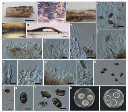
Saprobic on decaying bamboo culms. Sexual morph: Undetermined. Asexual morph: Stromata forming under a blackened area, up to 1–2mm long and 2–3 mm wide and becoming raised at maturity, ellipsoidal to irregular. Conidiomata 150–250 μm high, 500–800 μm diam., eustromatic, immersed in the stromata, globose to subglobose, dark brown, with ostiolate opening. Conidiomatal wall comprising two layers of cells of textura angularis, with dark to brown outer thin layer 5 μm thick, with 10–17 μm thick, hyaline, conidiogenous inner layer. Paraphyses 30–40 μm long × 2–3.5 μm wide, septate, wide at the base, curved. Conidiophores 5–12.5 × 2.5–4 μm (x = 7.4 × 3.6 μm, n = 20) short, hyaline, wide at base, smooth, formed from the inner cells of the conidiomata wall. Conidiogenous cells 2. 5–12 × 1.5– 3.5 μm (x = 8.1 × 2.5 μm, n = 20) obclavate, enteroblastic, phialidic, becoming annellidic, with 1–3 barely discernible annellations, indeterminate, discrete, cylindrical to ampulliform, hyaline, smooth-walled. Conidia 8.5– 10 × 4.5–5.5 μm (x = 9.5 × 5.2 μm, n = 20), ellipsoidal to oblong, occasionally obovoid, aseptate, straight, rounded at both ends, brown to dark brown, smooth-walled, with wall sparsely covered by small, roughened tubercules, inside usually containing 1–2 large guttules.
Culture characters: Conidia germinating on MEA within 24 h and germ tubes produced from sides. Colonies slow growing, 30 mm diam. after 15 days at 28–32 °C, under 12 h light/12 h dark, circular, with even margin, floccose at the center, drift white at margin, and light greenish brown at the centre.
Material examined: THAILAND, Chiang Rai Province, Mae Fah Luang Unversity, on dead culms of bamboo, 9 July 2013, Dong-Qin Dai, DDQ00261 (MFLU 15–1211, holotype); Ibid. (KUN HKAS88719, isotype), ex-type living cultures, MFLUCC 13–0854, ICMP 20529.
Notes: Roussoella tuberculata is characterized by immersed, eustromatic conidiomata, enteroblastic, phialidic, annellidic conidiogenous cells and ellipsoidal conidia covered by small tubercules. Roussoella tuberculata issimilar to R. hysterioides, but differs in having annellidic conidiogenous cells (Hyde et al. 1996). Conidia of Roussoella species usually haveverrucose wall ornamentation (Hyde et al. 1996; Liu et al. 2014), while R. tuberculata has conidia with small, roughened tubercules.
FIG. Roussoella tuberculata (MFLU 15–1211, holotype). a, b Conidiomata on bamboo culm. c Basal wall of conidioma. d, e Vertical sections of conidiomata. f, g, i–n Conidiogenous cells developing conidia (n: showing annellidic conidiogenous cells). h Paraphyses. p Germinating conidia. o, q, r Dark brown conidia (o, r: Mature conidia covered by small tubercules). s, t Cultures on MEA. Scale bars: a = 2 cm, b = 2 mm, b, d, e = 200 μm, c = 20 μm, f–j, m, o = 10 μm, k, l, n, p– r=5 μm