Resinoscypha variepilosa (R. Galán & Raitv.) T. Kosonen, Huhtinen & K. Hansen, comb. nov.
MycoBank number: MB 835740; Index Fungorum number: IF 835740; Facesoffungi number: FoF 14073; Fig. 14e, 20
Basionym. Protounguicularia variepilosa R. Galán & Raitv., Int. J. Mycol. Lichenol. 2: 223. 1986.
Synonym. Arachnopeziza variepilosa (R. Galán & Raitv.) Huhtinen, Mycotaxon 30: 14. 1987.
Holotype. SPAIN, Cádiz, Grazalema, Sierra del Pinar de Grazalema, 1200 m a.s.l., on dead wood of Abies pinsapo, 27 Nov. 1982, R. Galán (Herb. Galán 6117) !
Specimens examined. CANADA, Yukon, Kluane Lake, Outpost Mountain, on fallen, decayed trunk of Picea glauca, 19 Aug. 1987, S. Huhtinen 87/131 (TUR 99569). – DENMARK, Zealand, Sorø, Suserup forest reserve, on decorticated trunk of Fagus, 8 May 1994, J. HeilmannClausen 04-035 (TUR 124469). – SLOVAKIA, Bratislava Forest Park, along river Vydrica, on decorticated hardwood, 4 Aug. 1986, P. Lizon & S. Huhtinen 86/60 (TUR 92119). – SWEDEN, Upland, Haninge, Tyresta National Park, Bylsjöbäcken, on coniferous trunk, 1 June 2016, T. Kosonen & S. Huhtinen 16/41 (S, TUR). – UK, England, Hertfordshire, Wadesmill, on decayed wood of Ulmus, 24 Nov. 2010, K. Robinson (TUR 193603); Gloucestershire, Forest of Dean, Coalpit Hill, on a trunk of Betula pendula, 18 Apr. 2012, K. Robinson (TUR 196799).
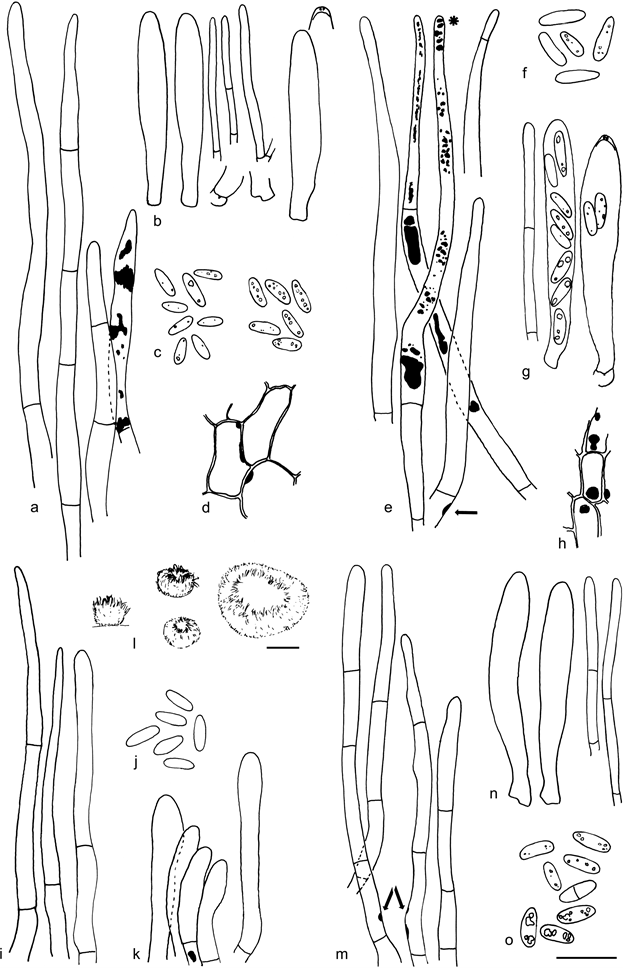
Notes — This is a distinct but rarely collected species with collections from two continents. Based on morphology alone it is difficult to assign a suitable genus to this species. However, in addition to the CB+ resin (Fig. 20a), also the very variable shape of (especially the shorter marginal) hairs is a characteristic feature, already shown by Raitviir & Galán (1986: f. 8 –11 of the holotype). The two collections, S. Huhtinen 87/131 and S. Huhtinen 16/41, from Canada and Sweden (Fig. 20e – k), have identical ITS and LSU sequences. The amount of resin on and in the hairs is highly variable even within one population, ranging from hairs practically without resin to hairs densely covered in resin (Fig. 20a, e, i). An ITS-LSU sequence of the Canadian sample has been previously deposited to GenBank (isolate M337 / collection S. Huhtinen 87/131: ITS, EU940163 and LSU, EU940086) (Stenroos et al. 2010). It was an erroneously annotated combination of two different species, the ITS presenting the correct species and the LSU apparently that of Pezoloma ciliifera of uncertain origin. The correct sequences were obtained by re-sequencing the original extraction of the isolate M337. The two sequenced samples were both from softwood.

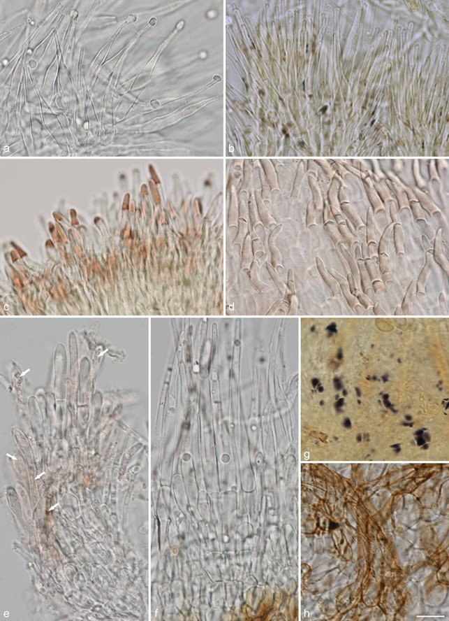
Fig. 14 Examples of hair shapes, inclusions and reactions, and of amyloid nodules in excipulum cells, in Hyaloscyphaceae. a. Thin-walled aseptate hairs,
Hyaloscypha spiralis (in water); b. hairs showing amyloid and dextrinoid nodules, Eupezizella aureliella (in MLZ); c– d. dextrinoid glassy hairs in Olla (in MLZ):
c. Olla transiens; d. Olla millepunctata; e. septate hairs showing resinous matter inside the hairs (arrows), Resinoscypha variepilosa (in water); f– h. Mimico scypha lacrimiformis: f. septate hairs with refractive globules; g. purple nodules in ectal excipulum cells (in MLZ); h. bulbous basal cells of hairs with exudates (in water) (a: KH.16.02; b: T. Kosonen 7296; c: U. Söderholm 4829; d: T. Kosonen 7155; e: S. Huhtinen 16/41; f– h. KH.17.02). — Scale bars = 10 µm. — All from fresh material. — Photos: a, e– h. K. Hansen; b– d. T. Kosonen.

Fig. 20 Resinoscypha variepilosa. a. Marginal hairs in CR, rightmost in CB showing the CB+ resinous contents; b. asci and paraphyses; c. spores in CB (left) and CR (right); d. ectal excipulum showing the amyloid nodules; e. marginal hairs showing the brown resinous substance inside, in MLZ, hair with asterisk (*) in water, one hair with basal amyloid nodule (arrow); f. spores in MLZ; g. asci and paraphysis; h. ectal excipulum showing the amyloid nodules; i. marginal hairs in CR, rightmost hair in water; j. spores in CR; k. short marginal hairs in MLZ showing one rare amyloid nodule; l. dry apothecia; m. marginal hairs in MLZ, some showing the amyloid nodules (arrows); n. asci and paraphyses; o. spores in CB, three lower in KOH (a – d: holotype; e– h: S. Huhtinen 87/131; i– k: S. Huhtinen 16/41; l– o: S. Huhtinen 86/60). — Scale bars: a– k, m– o = 10 µm, l = 100 µm. — Drawings: S. Huhtinen.
