Pseudotrichia rubriostiolata Phookamsak & K.D.Hyde.
Index Fungorum number: IF550933, Facesoffungi number: FoF00437; Fig. 1
Etymology – The specific epithet rubriostiolata refers to the ascomata ostioles with red pigments.
Holotypus – MFLU 11–0174
Saprobic on Thysanolaena maxima. Sexual morph Ascomata 140 – 220 μm high (excluding neck), 150 – 190 μm diam., solitary, scattered, immersed to semi-immersed or erumpent with neck, visible as dark spots, with reddish pigment surrounding the ostiole, uni – loculate, globose to subglobose, glabrous, ostiole central, with short papilla (70 – 95 × 70 – 90 μm). Peridium 5 – 20 μm wide, thin-walled, of equal thickness, composed of 2 – 3 layers, brown to dark brown, flattened, pseudoparenchymatous cells, arranged in a textura angularis. Hamathecium 1 – 1.5 (–2) μm wide, composed of dense, narrow cellular pseudoparaphyses, wall rough, with small guttules, distinctly septate, anastomosing above the asci, embedded in a hyaline gelatinous matrix. Asci (55–) 70 – 80 (–88) × (13–) 15 – 16 (–17) μm (x̄ = 75.5 × 15.4 μm, n = 25), 8 – spored, bitunicate, fissitunicate, clavate, short pedicellate with foot-like pedicel, apically rounded with well-developed ocular chamber. Ascospores (23.5–) 25 – 27 (–29) × 4 – 5 (–6) μm (x̄ = 26.5 × 4.5 μm, n = 30), overlapping 1 – 3-seriate, fusiform, slightly curved, hyaline, becoming light brown when geminated, 1 – septate, constricted at the septa, swollen near the septa, smooth-walled, surrounded by distinct mucilaginous sheath. Asexual morph forming stromatic fruiting bodies on bamboo pieces on water agar (WA) after 12 weeks. Conidiomata 80 – 205 μm high, 80 – 210 μm diam., pycnidial, scattered or clustered, gregarious, semi-immersed to superficial on bamboo pieces, visible as black spots on surface, globose to subglobose or irregular in shape, glabrous, surrounded by vegetative hyphae, ostiole undetectable, becoming reddish to reddish-purple. Conidiomata walls 7.5 – 15.5 μm wide, composed of several layers of dark brown to black, pseudoparenchymatous cells, initially arranged in textura intricata, becoming textura angularis. Conidiophores simple, rarely branched, aseptate, hyaline, mostly reduced to conidiogenous cells. Conidiogenous cells 4 – 16 × 1.5 – 2 (–3) μm (x̄ = 7×1.8μm, n=40), enteroblastic, phialidic, single, discrete, determinate, doliiform to cylindrical or ampulliform, hyaline, arising from basal stratum. Conidia 2 – 3.5 × 1 – 2 μm (x̄ = 3.1 × 1.6 μm, n = 40), one-celled, oblong or rod-shaped to obovoid, with rounded to obtuse ends, hyaline, smooth-walled.
Culture characters – Colonies on MEA slow growing, reaching 45 – 50 mm diam. after 4 weeks at 25 – 30 °C, cream to pale yellowish or pale reddish at the margin, white to cream or pale yellowish at the centre with small reddish droplets and the periphery of raised; reverse cream to white, yellowish or pale reddish at the margin, reddish to blond yellowish at the centre, slightly radiating; medium dense to dense, circular, flattened to slightly raised, dull with entire edge, initially mucoid, becoming velvety to felty, seperating from agar, produced reddish-brown to light brown pigments diffusing in the agar and conidiomata immersed in agar after 8 weeks.
Material examined – THAILAND, Chiang Rai Province, Muang District, Mae Fah Luang University campus grounds, on dead stem of Thysanolaena maxima (Poaceae), 13 August 2010, R. Phookamsak RP0054 (MFLU 11–0174, holotype), ex-type living culture, MFLUCC 11–0138, GenBank ITS:KP744463; SSU: KP744508; ibid., on dead stem of Thysanolaena maxima, 13 August2010, R. Phookamsak RP0053 (MFLU 11–0173), living culture, MFLUCC 11–0137. GenBank ITS: KP744462; LSU: KP744507; SSU:KP753966; Phrae Province, Rongkwang District, Maejo University Phrae Campus, on dead stem of Thysanolaena maxima, 20 August 2010, R. Phookamsak RP0072 (MFLU 11–0192), living culture, MFLUCC 11–0156. GenBank ITS:KP744464; LSU: KP744509; SSU: KP753967.
Notes – Pseudotrichia rubriostiolata clusters together with P. guatopoensis in the phylogenetic tree. Morphologically it differs from other Pseudotrichia species in the smaller size of its ascomata, asci and ascospores. P. rubriostiolata is similar to P. thailandica, but in P. rubriostiolata the ascomata, asci and ascospores are smaller and the ascospores are slightly constricted at the septum. In P. thailandica ascospores are larger and deeply constricted at the septum, and easily separate into two parts. In the phylogenetic analyses, P. rubriostiolata forms a clade with P. thailandica and P.guatopoensis (strain SMH 4535).
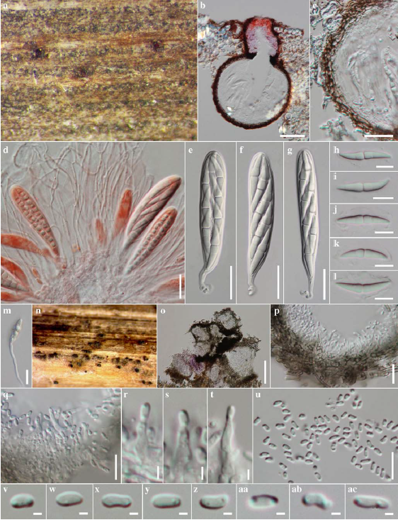
Fig. 1 Pseudotrichia rubriostiolata (holotype) a Ascomata on host surface visible as black raised areas with reddish pigments at the apex b Section through an ascoma c Section through peridium d Asci with pseudoparaphyses stained in Congo red reagent e – g Asci h – l Ascospores m Germinating spore n Pycnidia on bamboo pieces on WA o Section through stromatic pycnidia p Section though pycnidial walls q – t Conidiophores u – ac Conidia. Scale bars: b = 50 μm, c – g, m = 20 μm, h – l, p – u =1 0μm, r – t = 2 μm, v – ac = 1 μm, o = 100 μm.