Pseudotrichia thailandica Phookamsak & K.D. Hyde.
Index Fungorum number: IF550934, Facesoffungi number: FoF 00438; Fig. 2
Etymology – The specific epithet thailandica refer to the country in which the fungus was first collected.
Holotypus – MFLU 11–0191
Saprobic on dead stem of Thysanolaena maxima. Sexual morph Ascomata 140 – 180 μm high (including neck), 165 – 310 μm diam., solitary to gregarious, scattered, semi-immersed, visible on host surface as raised, dark spots, with reddish pigment surrounding the ostiole, uni-loculate, rarely bi-loculate, subglobose, glabrous, ostiole oblique, with short papilla, carbonaceous. Peridium 7 – 22 μm wide, thin-walled, of unequal thickness, slightly thin at the base of ascoma, composed of several layers of brown to dark brown, pseudoparenchymatous cells, outer layer comprising 2–3 layers, flattened, arranged in a textura prismatica, inner layer comprising 2 – 3 layers, arranged in textura angularis. Hamathecium composed of dense, 0.8–2.5μm wide, dense, narrow, cellular pseudoparaphyses, wall rough with small guttules, distinctly septate, anastomosing above asci, embedded in a hyaline gelatinous matrix. Asci (77.5–) 80 – 100 (–115.5) × (14–) 16 – 18 (–20) μm (x̄ = 95.1 × 17.3 μm, n = 25), 8 – spored, bitunicate, fissitunicate, clavate, short pedicellate with foot-like or knob-like pedicel, apically rounded, with well-developed ocular chamber. Ascospores (27–) 28 – 32 (–34.5) × 4 – 5 (–6) μm (x̄ = 30 × 4.7 μm, n = 30), overlapping 1 – 3 – seriate, fusiform, slightly curved, hyaline to subhyaline, 1 (–5)-septate, constricted at the septa, easily seperated into two part spores, swollen near the septa in the upper cell, smooth-walled, surrounded by a distinct mucilaginous sheath. Asexual morph Undetermined.
Culture characters – Colonies on PDA slow growing, reaching 30 – 35 mm diam. after 4 weeks at 25 – 30 °C, cream to pale yellowish at the margin, grey to dark grey at the centre, with small black droplets; reverse cream to yellowish-white at the margin, dark greenish at the centre, slighty radiating, forming sectors; medium dense to dense, circular, flattened to slightly raised, dull with undulate edge, mucoid, fairly fluffy at the magins, seperating from agar, felty in the centre with sparse hyphae at the margin.
Material examined – THAILAND: Phrae Province, Rongkwang District, Maejo University Phrae Campus, on dead stem of Thysanolaena maxima (Poaceae), 20 August 2010, R. Phookamsak RP0071 (MFLU 11–0191, holotype), ex-type living culture, MFLUCC 11–0155.
GenBank Accession numbers – ITS:KP744465; LSU: KP744510; SSU: KP753968.
Notes – Pseudotrichia thailandica is similar to P. rubriostiolata in having a red pigment in the ostiole. However, Pseudotrichia thailandica has larger ascomata, asci and ascospores than P. rubriostiolata. These two species form a clade together with P. guatopoensis (strain SMH 4535) in the phylogenetic tree (Fig. 1).
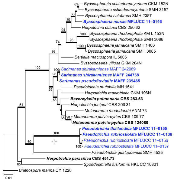
Fig. 1 Phylogram generated from Maximum likelihood (RAxML) analysis based on combined LSU, SSU and TEF1 sequence data of Melannomataceae. Maximum likelihood bootstrap support values greater than 50 % are indicated above or below the nodes, and branches with Bayesian posterior probabilities greater than 0.95 are given in bold. The ex-types (reference strains) are in bold; the new isolates are in blue. The tree is rooted with Biatriospora marina strain CY 1228.
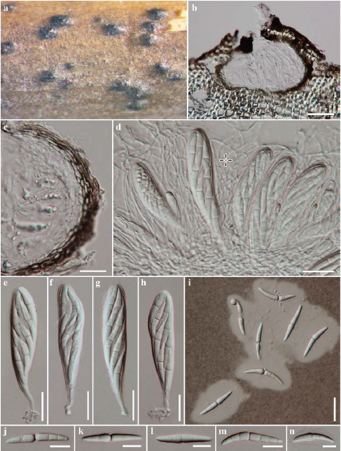
Fig. 2 Pseudotrichia thailandica (holotype) a Ascomata on host surface b Section through an ascoma c Peridium d Asci with pseudoparaphyses e – h Asci i Ascospores in Indian ink. j – n Ascospores. Scale bars: b = 50 μm, c – i = 20 μm, j – n = 10 μm, o = 100 μm.