Byssosphaeria musae Phookamsak & K.D. Hyde.
Index Fungorum number: IF550932, Facesoffungi number: FoF00436; Fig. 2
Etymology – The specific epithet musae refers to the host.
Holotypus – MFLU 11–0182
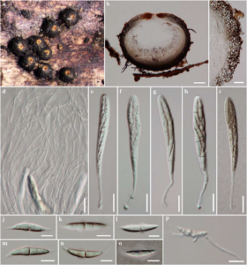
Saprobic on leaf sheath of Musa sp. Sexual morph Ascomata 430 – 540 μm high, 450 – 630 μm diam., gregarious, scattered, superficial on subiculum, visible as dark spots on host, orange to yellow around pore, uni-loculate, globose to subglobose, setose, apex rounded, ostiole central, with pore-like opening. Peridium 35 – 80 μm wide, thick-walled, of equal thickness, composed of several layers of dark brown to black cells, arranged in textura angularis to textura prismatica. Hamathecium 0.5 – 1.7 μm wide, composed of dense, trabeculate, distinctly septate, anastomosing, pseudoparaphyses, embedded in a hyaline gelatinous matrix. Asci (120–) 125 – 135 (–145) × (11.5–) 12 – 14 (–17) μm (x̄ = 132.8 × 13.7 μm, n = 25), 8 – spored, bitunicate, fissitunicate, clavate, long pedicellate (40–50μm long) with knob-like pedicel, apically rounded, with well-developed ocular chamber. Ascospores (27–) 30 – 33 (–36) × (4–) 5 – 6 μm (x̄ = 32.3 × 5.9 μm, n = 30), overlapping 1 – 2 – seriate, fusiform, with acute ends, hyaline to pale brown when young, becoming light brown at maturity, 1 (–3)-septate, not constricted at the septa, slightly curved, smoothwalled, bearing delicate hyaline appendages over ends with wing-like appendages near the central septum. Asexual morph Undetermined.
Culture characters – Colonies on PDA fast growing, 80 – 90 mm diam. after 2 weeks at 2 – 30 °C, white to cream or pale yellowish, intermixed with yellowish to orangish hyphae; reverse cream to white yellowish at the margin, yellowish-brown at the centre, medium dense to dense, irregular, flattened to slightly raised, dull with entire edge, fluffy to feathery, effuse, zonate.
Material examined – THAILAND: Chiang Rai, Muang District, Khun Korn Waterfall, on leaf sheath of Musa sp. (Musaceae), 5 September 2010, R. Phookamsak RP0062 (MFLU 11–0182, holotype); ex-type living culture, MFLUCC 11–0146. GenBank ITS: KP744435; LSU: KP744477; SSU: KP753947.
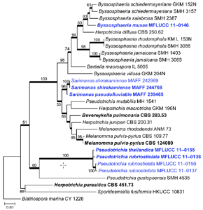
Notes – Byssosphaeria musae is similar to B. salebrosa (Cooke & Peck) M.E. Barr and B. schiedermayeriana (Fuckel) M.E. Barr (1984; Mugambi and Huhndorf 2009), with ascomata, asci and ascospores having a similar size range, but being smallest in B. musae which has smaller ascomata, asci and ascospores than these species. Byssosphaeria musae differs from B. schiedermayeriana in ascospore appendages, which are present in B. musae, but lacking in B. schiedermayeriana. Phylogenetic analyses of combined genes (Fig. 1) showed that Byssosphaeria musae forms a separate robust clade (77 % MP and 67 % ML) close to B. schiedermayeriana and B. salebrosa.

Fig. 1 Phylogram generated from Maximum likelihood (RAxML) analysis based on combined LSU, SSU and TEF1 sequence data of Melannomataceae. Maximum likelihood bootstrap support values greater than 50 % are indicated above or below the nodes, and branches with Bayesian posterior probabilities greater than 0.95 are given in bold. The ex-types (reference strains) are in bold; the new isolates are in blue. The tree is rooted with Biatriospora marina strain CY 1228.

Fig. 2 Byssosphaeria musae (holotype) a Ascomata on host surface b Section through ascoma c Peridium d Pseudoparaphyses e–i Asci j–n Ascospores o Ascospore stained in Indian ink. p Germinating spore. Scale bars: b = 100 μm, c – i, p = 20 μm, j – o = 10 μm.
