Phyllosticta citribrasiliensis O.L. Pereira, Glienke & Crous [as ‘citribraziliensis’], in Glienke, Pereira, Stringari, Fabris, Kava-Cordeiro, Galli-Terasawa, Cunnington, Shivas, Groenewald & Crous, Persoonia 26: 54 (2011) Fig. 1
MycoBank number: MB 517971; Index Fungorum number: IF 831482; Facesoffungi number: FoF 12964;
Saprobic on Laburnum sp. Sexual morph Not observed. Asexual morph Coelomycetous. Conidiomata 100–160 × 80–110 µm (x̄=111 × 97.5 μm, n=15), solitary, uniloculate, globose to sub-globose, scattered, semi-immersed, conspicuous on host surface, black. Pycnidial walls 13.7–27.0 μm wide (x̄=18.6 μm, n=15), comprising several layers of textura angularis cells, outer layers dark brown to black, inner layers pale brown to hyaline. Ostiole single, central, 12.5–17.5 μm wide (x̄=14.8 μm, n=5). Conidiophores reduced to conidiogenous cells. Conidiogenous cells 5.60–15.0 × 1.30–2.60 μm (x̄=10.1 × 2.28 µm, n=15), enteroblastic, phialidic, integrated, truncate to cylindrical to ampulliform, hyaline, formed from the inner layer of pycnidial wall. Conidia 7.0–9.8 × 4.5–6.6 µm (x̄=8.5 × 5.72 μm, n=30), solitary, ellipsoidal to obovoid, coarsely guttulate, aseptate, hyaline, smooth-walled, with narrowly truncate base, surrounded by a mucilaginous sheath, thicker on both sides, 1.3–2.0 µm thick (x̄=1.58 μm, n=20), thinner at the apex and base, 0.35–0.90 µm thick (x̄=0.64 μm, n=20), and bearing a hyaline, apical mucoid appendage. Appendage 2.85–27.0 × 0.70–1.80 µm (x̄=8.30 × 1.09 µm, n=20), flexible, unbranched, tapering towards an acutely rounded tip.
Material examined – Russia, on dead leaves of Laburnum sp., 15 October 2018, Timur S. Bulgakov T-7583 (MFLU XXX).
Distribution – Brazil, Russia (This study) (Farr and Rossman 2022).
Notes – Our strain clusters with the type strain (CBS 100098T) as well as other strains of P. citribrasiliensis (LGMF09, LGMF08, CPC 17466, CPC 17465, CPC17464) with strong support (87%ML) (Figure 2). In pairwise nucleotide comparisons of the type species of P. citribrasiliensis and our isolate, there are 0 nucleotide base pair (bp) differences in LSU (759 nucleotides), ef1α (207 nucleotides), and act (215 nucleotides). Morphological differences and similarities are given (Table 7). Coupled with multigene phylogenetic analyses and morphological characters, we establish our strain as a new host and country record. This is the first time we report P. citribrasiliensis from Laburnum sp. in Russia (Farr and Rossman 2022).
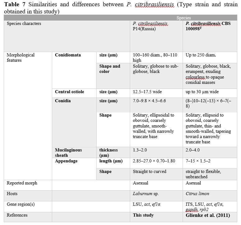
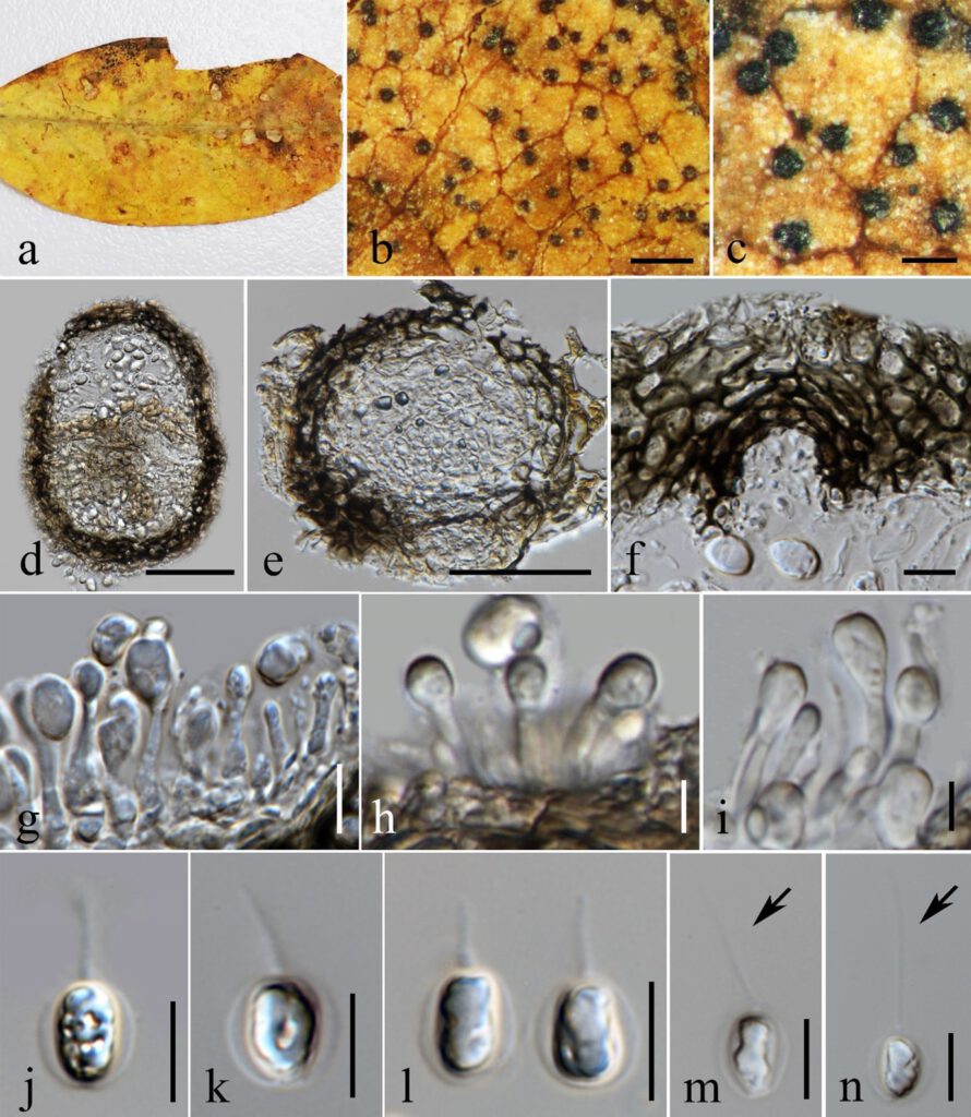
Figure 1 – Phyllosticta citribrasiliensis (MFLU XXX) a–b Appearance of conidiomata on leaves of Laburnum sp. c Close up of conidiomata on substrate. d–e Section through conidiomata showing pycnidial wall. f Ostiole g–i Development of conidia from phialides. j–n Conidia surrounded by mucilaginous sheath, bearing an apical appendage. Scale bars: b = 500 μm c = 200 μm, d–e = 50 μm, f, g, j–n = 10 μm, h–i = 5 μm.
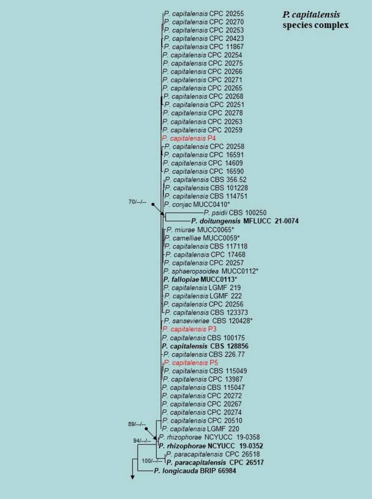
Figure 2 – Phylogram generated from maximum likelihood analysis (RAxML) based on the combined ITS, LSU, EF-1α, ACT, GAPDH and RPB2 matrices of Phyllosticta. Maximum likelihood (ML) with bootstrap support ≥60% is given at respective nodes. The tree is rooted with Botryosphaeria obtusa (CMW 8232 and CMW 7775) and B. stevensii (CBS 112553 and CMW 7060). Ex-type strains are indicated in bold and our isolates are in bold red.
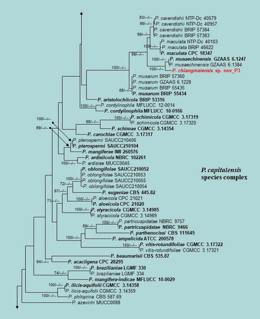
Figure 2 – Continued.
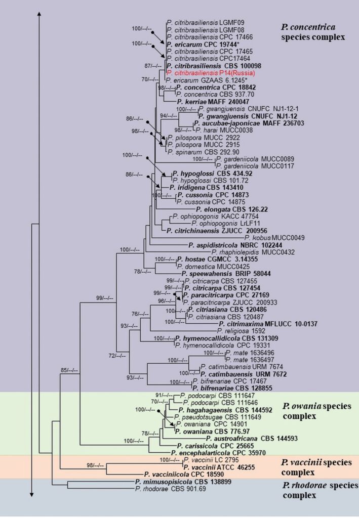
Figure 2 – Continued.
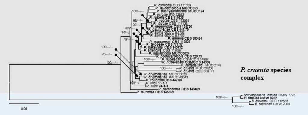
Figure 2 – Continued.
