Neomicrosphaeropsis tamaricicola (Wanas., Camporesi, E.B.G. Jones & K.D. Hyde) Thambugala, Wanasinghe & K.D. Hyde, comb. nov. Index Fungorum Number: IF552093
Basionym: Phoma tamaricicola Wanas., Camporesi, E.B.G. Jones & K.D. Hyde, in Crous et al., Persoonia, Mol. Phyl. Evol. Fungi 33: 281 (2014).
Saprobic or weak pathogen on twigs and branches of Tamarix species. Sexual morph: Ascomata solitary, scattered, immersed, slightly erumpent, dark brown to black, ostiolate. Peridium comprising 6–8 layers, outer layers heavily pigmented, thick-walled, comprising blackish to dark brown cells of textura angularis, inner layers composed of lightly pigmented to hyaline thin-walled cells of textura angularis. Hamathecium comprising numerous, filamentous, branched, septate pseudoparaphyses. Asci 8-spored, bitunicate, fissitunicate, cylindrical to cylindric-clavate, pedicellate, apically rounded, with a minute ocular chamber. Ascospores 1–2-seriate, partially overlapping, muriform, ellipsoidal, 4–6 transversely septate, with 3–4 vertical septa, constricted at the central septum, initially hyaline, becoming yellowish brown at maturity, conical and narrowly rounded at the ends, smooth-walled, without a mucilaginous sheath (description modified from Crous et al. 2014). Asexual morph: Conidiomata 65–155 μm diam. × 75–120 μm high (x̅ = 115 × 97 μm, n = 6), scattered to gregarious, immersed to erumpent, black, subglobose, uniloculate, ostiolate. Conidiomatal wall 12–22 μm composed of 3–5 layers of dark brown, thick-walled cells of textura angularis, fusing at the outside with the host tissues. Conidiogenous cells 2–4 × 1.6–3.2 μm (x̅= 3.1 × 2.2 μm, n = 15), enteroblastic, phialidic, hyaline, cylindrical, discrete or integrated, smooth. Conidia 3.5–6.6 × 2.5–3.4 μm (x ̅= 5.3 × 2.9 μm, n = 40), ellipsoidal or obovoid, hyaline to light brown, straight, rounded at both ends, 1-celled, smooth-walled.
Culture characteristics: Conidia germinating on PDA within 18 h. Colonies growing on PDA reaching 30 mm diam. After 10 days at 25 °C, circular, flat, moderately dense, surface grey, olivaceous brown to dull green from below, surface smooth with entire to slightly undulate edge.
Material examined: ITALY, Province of Forlì-Cesena, Ravaldino in Monte – Forlì, on dead branches of Tamarix gallica L. (Tamaricaceae), 15 January 2014, Erio Camporesi (MFLU 14–0333, holotype); ibid. 28 March 2014, Erio Camporesi IT 1785C (MFLU 14–0602), living culture MFLUCC 14–0443, ICMP 20708; ibid. 22 November 2012 Erio Camporesi IT 918–1 (MFLU 14–0591), living culture MFLUCC 14–0439, ICMP 20743.
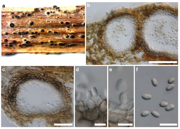
FIG Neomicrosphaeropsis tamaricicola (MFLU 14–0602, asexual morph) a Conidiomata on host surface b, c Vertical sections through conidiomata d, e Conidiogenous cells and developing conidia f conidia. Scale bars: b = 50 μm, c = 30 μm, d-e = 5 μm, f = 10 μm