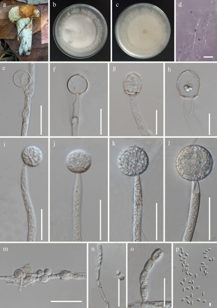
Mucor nederlandicus Váňová, Česká Mykol. 45(3): 128 (1991) Figure 6
MycoBank number: MB 354494; Index Fungorum number: IF 354494; Facesoffungi number: FoF 12295;
Fungicolous on a fruiting body of Russula sp. Asexual morph on PDA: Sporangiophores up to 3.2–9.5 μm (x̅ = 6.5 μm, n = 20) width, hyaline, undulate, occasionally curved, irregular septate near the base, unbranched, sympodial branches formed. Sporangia 19.5–38.5 × 18.5–35.5 μm (x̅ = 29 × 27 μm, n = 10), globose, thick-walled, wall echinulate, deliquescent in mature sporangia, hyaline to pale brown. Columellae 14.7–23.5 × 14.5–23 μm (x̅ = 19 × 18.5 μm, n = 20), mostly globose to subglobose, and sometimes oblong, obovoid, and rarely ellipsoid, with or without short collar, hyaline to pale brown, smooth-walled. Sporangiospores 3.5–6.5 × 2.2–3.5 μm (x̅ = 5 × 3 μm, n = 45), mostly ellipsoidal, sometimes flattened on one side, oval or cylindrical, smooth-walled, hyaline with one or more granules. Chlamydospores abundant, intercalary, terminal, variable in shape and size. Rhizoids absent. Sexual morph: not observed.
Culture characteristics: on PDA, at 25 °C, cultures are cottony and white to pale yellow. The colony reached 60 mm in diam. after 4 days of incubation.
Materials examined: Thailand, Chiang Mai Province, Mae Tang, Pa Pae, Ban Pha Deng, isolated from the white mycelium growing on a fruiting body of Russula sp., 07 July 2021, Gajanayake AJ, MR 30 inactive dry culture MFLU aaaa, living cultures MFLUCC aaaa and MFLUCC bbbb; Thailand, Chiang Mai province, Mae Tang, Pa Pae, Ban Pha Deng, isolated from the white mycelium growing on a fruiting body of Russula sp., 07 July 2021, Gajanayake AJ, MR 33 inactive dry culture MFLU xxxx, live cultures MFLUCC xxxx and MFLUCC xxxx.
GenBank accession numbers: (MFLUCC aaaa)- ITS: xxx, LSU: xxx; (MFLUCC bbbb)- ITS: xxx, LSU: xxx
Notes: Mucor nederlandicus strains MFLUCC aaaa and MFLUCC bbbb reported in this study share similar asexual morphologies with those described by Oudem 1898 as mentioned in Mycobank (2022). The sporangia, sporangiospores and columellae of MFLUCC aaaa and MFLUCC bbbb are comparatively smaller than M. nederlandicus described by Oudem 1898 as mentioned in Mycobank (https://www.mycobank.org/), as sporangia; 18.5–35.5 μm vs 40–45 µm in diam., sporangiospores; 5 × 3 μm vs 7 × 3 µm, columellae; 14.5–23 μm vs 25–35 µm in diam. These dimensional differences might have occurred due to the variations of agar medium and the conditions used to obtain the culture. To the best of our knowledge, this is the first report of M. nederlandicus on fruiting bodies of Russula sp. as a fungicolous fungus.

Figure 6: Mucor nederlandicus (xxx) a Host b & c Colony on PDA d Sporulation on PDA e-h Columella i-l Sporangia m-o Chlamydospores p Sporangiospores. Scale bars: d = 500 μm, l, n = 50 μm, i-k,m,o = 30 μm, e = 20 μm, f-h = 15 μm, p = 5 μm.
