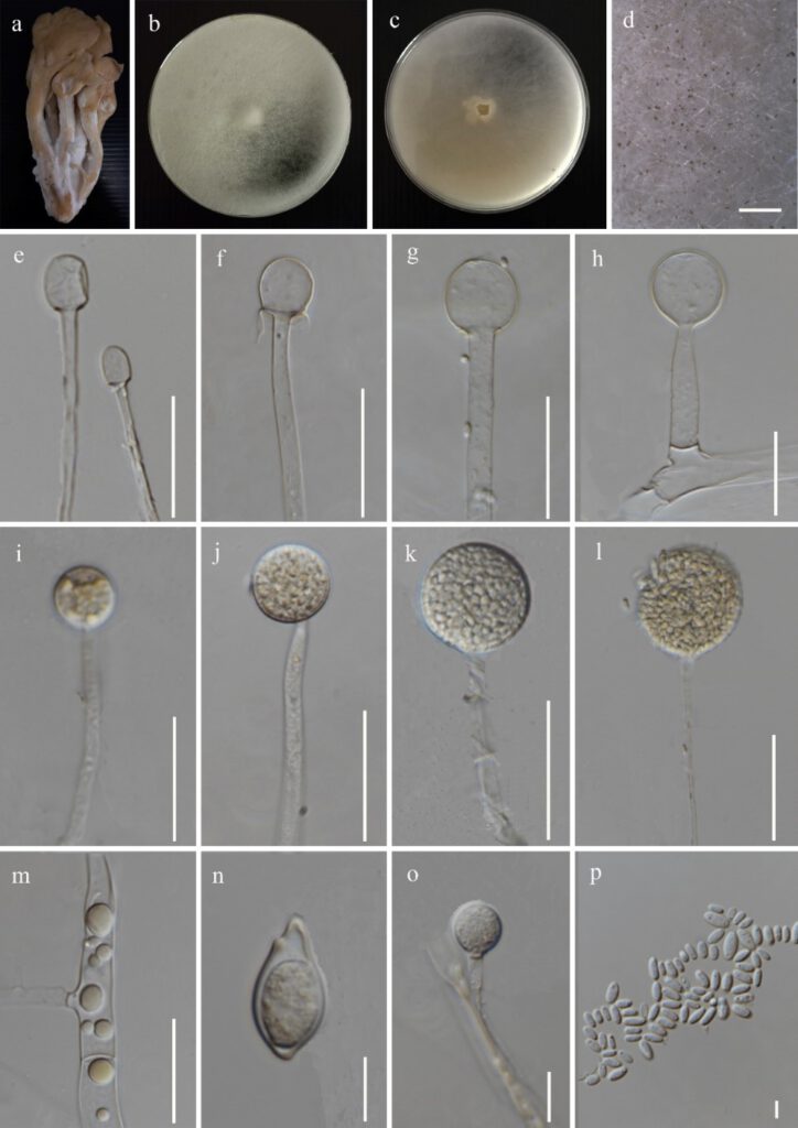
Mucor irregularis Stchigel, Cano, Guarro & E. Álvarez, in Álvarez et al., Medical Mycol. 49(1): 71 (2011) Figure 5
MycoBank number: MB 546922; Index Fungorum number: IF 546922; Facesoffungi number: FoF 12294;
Fungicolous on a fruiting body of Pleurotus sp. Asexual morph on PDA: Sporangiophores up to 3.8–10.5 μm (x̅ = 7.2 μm, n = 20) width, hyaline, septate, erect, sympodially and monopodially branched. Sporangia 20.5–38.5 × 20–38 μm (x̅ = 29.5 × 29 μm, n = 10), globose to sub-globose, grayish-brown, with diffluent wall. Columellae 13.5–30 × 12.5–29 μm (x̅ = 22 × 20.5 μm, n = 20), globose or ellipsoidal to cylindrical, rarely conical, smooth-walled, have small or no clear collars. Sporangiospores 3–7 × 1.5–3 μm (x̅ = 4.5 × 2 μm, n = 45), hyaline, smooth-walled, mostly ellipsoidal to ovoid, or globose, some irregularly shaped. Rhizoids not observed. Chlamydospores vary in shape and size. Sexual morph: not observed.
Culture characteristics: on PDA, at 25 °C, cultures are initially white and later turn yellowish with sporangia formation. The colony reached 84 mm in diam. after 4 days of incubation.
Materials examined: Thailand, Chiang Rai Province, Muang, Ban Du, isolated from the black patches in white mycelium grown on a fruiting body of Pleurotus sp., 28 October 2021, Gajanayake AJ, AJ 010 inactive dry culture MFLU yyy, living culture MFLUCC yyy.
GenBank accession numbers: ITS: xxx, LSU: xxx
Notes: The morphological characteristics of Mucor irregularis (MFLU yyy) described here show similarity to the original description given by Zheng and Chen (1991), as Rhizomucor variabilis which has later been synonymized to M. irregularis in Alvarez et al. (2011). The sporangia, sporangiospores and columellae MFLUCC yyy are comparatively smaller than M. irregularis, described by Zheng and Chen (1991) as sporangia; 20–38 μm vs (23.5–) 35–106 μm in diam., sporangiospores; 3–7 × 1.5–3 μm vs 2.5–11.5 (–16.5) × 2–7 μm and columellae; 13.5–30 × 12.5–29 μm vs 23.5– 71× 26–82.5 μm. In contrast to M. irregularis described in Zheng and Chen (1991), highly variable shapes of columellae and sporangiospores were not observed in MFLUCC yyy. These dimensional and shape differences may have been occurred due to the variations of agar medium and the conditions used to obtain the cultures. To the best of our knowledge, this is the first report of M. irregularis on a fruiting body of Pleurotus sp. as a fungicolous fungus.

Figure 5: Mucor irregularis (xxx) a Host b & c Colony on PDA d Sporulation on PDA e-h Columella with and without collars i-l Sporangia m Granular content inside hyphae n & o Chlamydospores p Sporangiospores. Scale bars: d = 200 μm, e-g, j-m = 30 μm, h & i = 20 μm, n,o = 10 μm, p = 5 μm.
