Phacidiella kunmingensis D.P. Wei and K.D. Hyde, sp. nov. Fig. 2
MycoBank number: MB; Index Fungorum number: IF; Facesoffungi number: FoF 12296;
Etymology: The specific epithet is derived from Kunming County, Yunnan Province, China
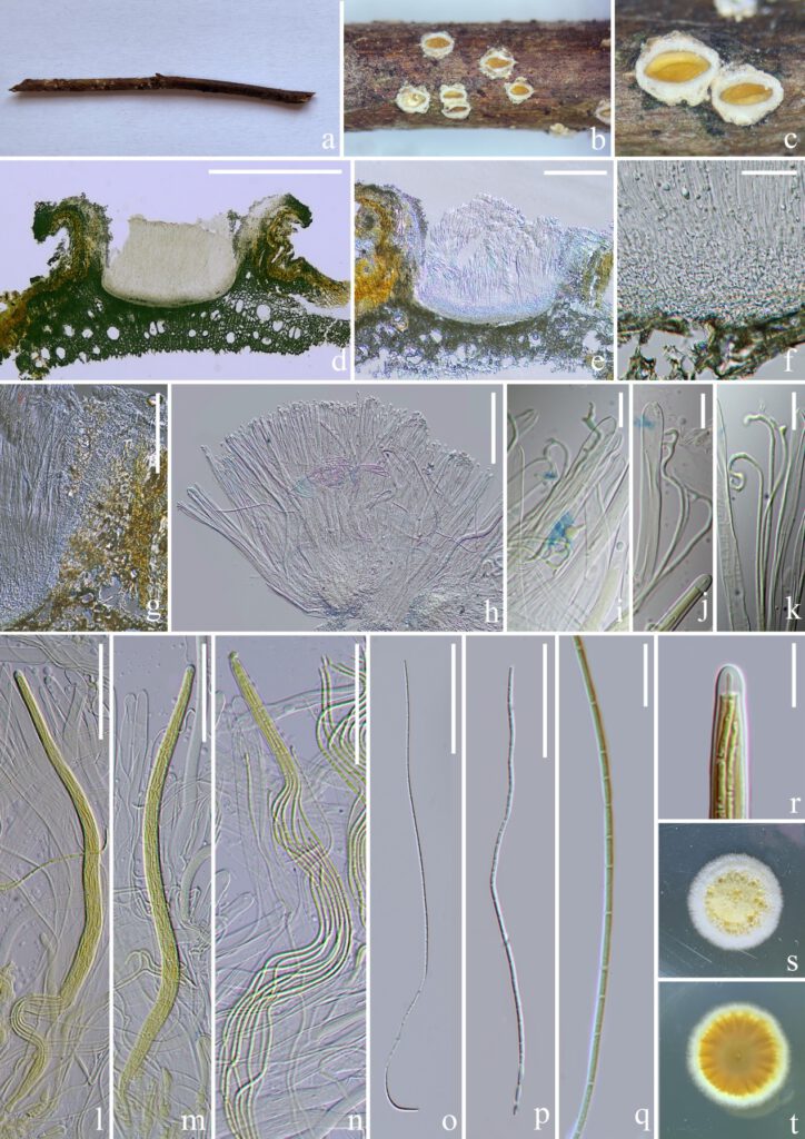
Saprobic on dead twigs. Sexual morph: Apothecia 400–650 × 350–450 (x̄ = 510× 407, n = 5) μm, clustered, immersed, cupulate, opening by a finely large pore. Margin entire to lacerate, white-pruinose, turnup outward. Disc deeply immersed, yellow, splitting away from the margin when dry. Exciple 50–120 (x̄ = 85, n = 30) μm, four-layered, with 1) an prominent accessory thalline margin, 2) a wall extended from subhymenium, of hyaline, thick-walled cell of textura angularis, 3) a crystalliferous layer where crystals are abundant particularly in the upper part and 4) a compact periphysoidal layer (10–17 (x̄ = 13, n = 20) μm) perpendicularly lined with the crystalliferous layer. Subhymenium 30–45 (x̄ = 36, n = 30) μm, of hyaline, angular cells, J–. Paraphyses 1–2 (x̄ = 1.5, n = 35) μm in width, filiform, numerous, aseptate, apically branched, enlarged, circinate, faintly J+, not gelatinous, as long as asci. Marginal paraphyses not observed. Asci 200–260 × 4.5–7.5 (x̄ = 233 × 5.8, n = 25) μm, eight-spored, cylindrical, slightly tapering toward the base, thick-walled, with a thick apex. Ascus cap 3.5–5 × 3–5 (x̄ = 4.2 × 3.9, n = 20) μm, round, pierced by a pore. Ascospores 125–230 × 1–1.8 (x̄ = 184 × 1.3, n = 20) μm, filiform, multi-septate, flexuous, non-disarticulating, no contraction at septa, the interval cell 4.8–10 (x̄ = 6.6, n = 35) μm in length. Asexual morph: Undetermined.
Culture characteristics: Spores were germinated at 25 ℃ after two days. Colonies on PDA growing slowly, reaching 2.2 cm in diameter after 53 days. The colony was pale creamy yellow at center and white at the periphery. It was circular, cotton, slightly raised, with an entire margin and dense mycelia, reverse yellow, with radiation stripes. No pigments were produced in agar.
Material examined: China, Yunnan Province, Kunming City, Panlong district, near Songhuaba reservoir, 11 December 2021, on dead twigs of an unidentified deciduous host, Deping Wei, SHB1226a (HKAS 124175, holotype), (KUNCC 22-10811, ex-type living culture), ibid. SHB1226b (HKAS 124176), (KUNCC 22-10812, living culture).
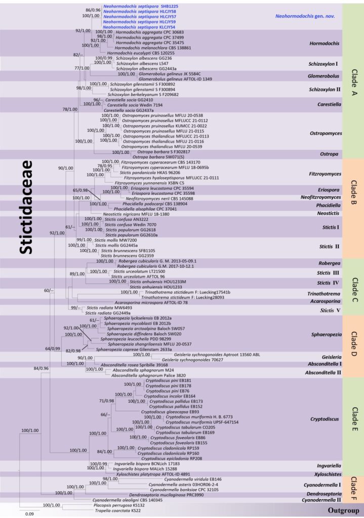
Notes: BLASTn results (accessed on 18 May 2022) of the LSU sequence of Phacidiella kunmingensis revealed 95.5% similarity to P. alsophilae (accession no. MT373344), 94% to P. podocarpi (accession no. NG_058118) and 96% to Fitzroyomyces yunnanensis (accession no. MZ781317). The ITS sequence revealed 88.35% similarity to P. alsophilae (accession no. MT373361), 84.68% to P. podocarpi (accession no. KP004453), and 82.11% to Fitzroyomyces cyperacearum (accession no. MK499349). The mtSSU sequence revealed 92.06% to Fitzroyomyces hyaloseptisporus (accession no.MZ868911), 92.21% to F. yunnanensis (accession no. MZ781329) and 88.89% to Carestiella socia (accession no. AY661678). In the phylogenetic tree, Phacidiella kunmingensis is sister to P. alsophilae and P. podocarpi with strong support (98% MLBS/ 100% PP, Figure 1). It is impossible to compare the morphology of P. kunmingensis with known Phacidiella species because the latter are known exclusively from their asexual morphs. A nucleotide comparison between P. kunmingensis and P. alsophilae shows that there are 69 bp differences (including 32 gaps) and 86 bp differences (including 27 gaps) across 1003 bp of LSU and 735 bp of ITS region, respectively. Phacidiella kunmingensis differs from P. podocarpi in 90 bp (including 51 gaps) in the LSU (1035 bp) and 139 bp (including 38 gaps) in the ITS (820 bp).

Figure 1. Phylogram generated from a maximum likelihood analysis based on concatenated ITS, LSU, mtSSU and RPB2 sequence datasets. Bootstrap support (MLBS) equal or greater than 60% and Bayesian posterior probabilities (PP) equal or higher than 0.95 are given above the nodes. Generic names are noted in the right side. Newly generated sequence are highlighted in blue bold font.

Fig. 2 Phacidiella kunmingensis. (holotype, SHB1226a). a Substrate. b, c Apothecia. d, e Vertical section of apothecia. f Subhymenium. g Exciplum. h Hymenium. i–k Paraphyses showing faintly blue iodine reaction. l–m Asci. o–q Ascospores. r Ascus cap. s, t Upper and lower view of culture on PDA after 48 days. Scale bars: d = 500 µm, e = 200 µm, g, h = 100 µm, l–p = 50 µm, f = 30 µm, i–k, q, r = 10 µm. (i–n, r were treated with Melzer’s reagent)
