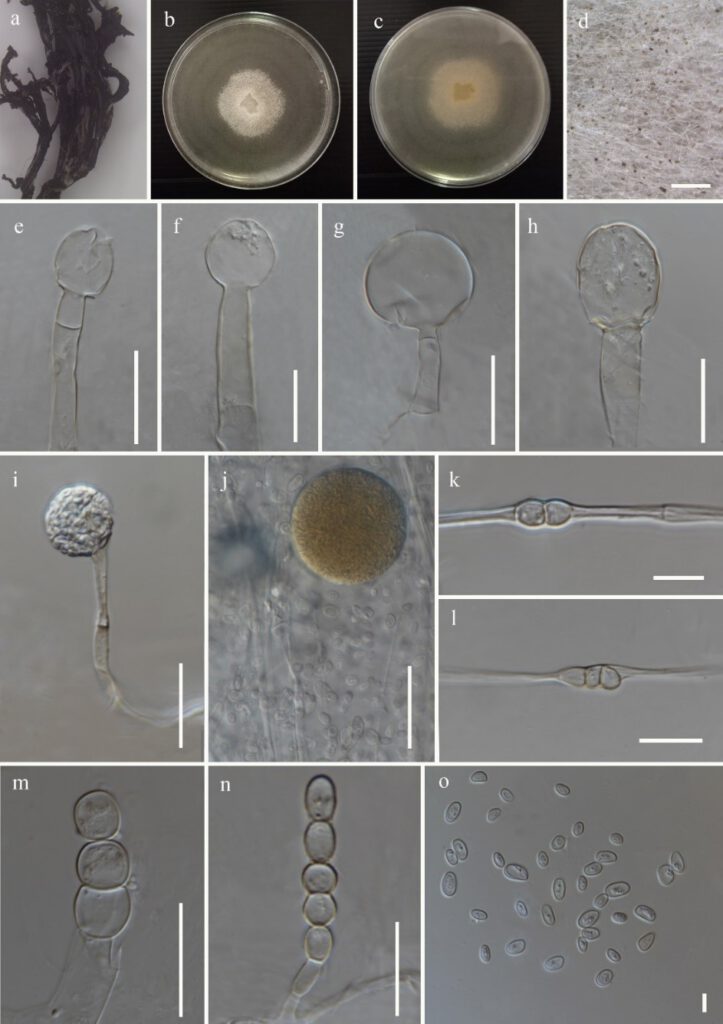
Mucor hiemalis Wehmer, Annls mycol. 1(1): 39 (1903) Figure 4
MycoBank number: MB 249401; Index Fungorum number: IF 249401; Facesoffungi number: FoF 12293;
Fungicolous on a fruiting body of Ramaria sp. Asexual morph on PDA: Sporangiophores up to 12–23 μm (x̅ = 12.5 μm, n = 20) width, slightly branched sympodially, septate. Sporangia 42–45 × 40–43 μm (x̅ = 43.5 × 41.5 μm, n = 05) globose, initially hyaline to yellowish, then dark brown when matured, with deliquescent, slightly transparent walls. Columellae 28–38.5 × 26.5–30.5 μm (x̅ = 33 × 30 μm, n = 20) ellipsoidal with truncate base, globose when young. Sporangiospores 3.7–7.8 × 2.5–5 μm (x̅ = 3.5 × 5.5 μm, n = 45) ellipsoidal, containing a few scattered granules, variable in size. Chlamydospores intercalary, terminal, variable in shape and size. Sexual morph not observed.
Culture characteristics: on PDA, 25 °C, cultures are initially white and later turn yellowish. The colony reached 50 mm diam. after 4 days of incubation.
Materials examined: China, Yunnan Province, Lijiang, Shigu, isolated from the white mycelium growing on a fruiting body of Ramaria sp., 29 June 2020, Karunarathna SC, SG 039, Herbarium material ZHKU 22-0059, living culture ZHKUCC 22-0111; China, Yunnan Province, Lijiang, Shigu, isolated from the white mycelium growing on a fruiting body of Boletus sp., 29 June 2020, Karunarathna SC, SG 071, Herbarium material ZHKU 22-0060, living culture ZHKUCC 22-0112.
GenBank accession numbers: ZHKUCC 22-0111- ITS: xxx, LSU: xxx, ZHKUCC 22-0112- ITS: xxx, LSU: xxx
Notes: Mucor hiemalis strains ZHKUCC 22-0111 and ZHKUCC 22–0112 reported in this study share similar asexual morphologies with those observed from the ex-neotype (CBS 201.65) of M. hiemalis described by Schipper (1973). During the micro-morphologcal examination, only 05 sporangia were measured as most of the sporangia were easily broken during the slide preparation due to the prevalence of deliquescent sporangial wall. The sporangia, sporangiospores and columellae of ZHKUCC 22–0111 and ZHKUCC 22-0112 are comparatively smaller than M. hiemalis described by Schipper (1973) as sporangia; 41.5 µm vs 70 µm in width, sporangiospores; 3.7–7.8 × 2.5–5 μm vs 5.7–8.7 × 2.7–5.4 µm and columellae; 33 × 30 μm vs 38 × 30 µm. The sporangiophores of ZHKUCC 22–0111 and ZHKUCC 22–0112 (up to 12–23 μm) are comparatively bigger than M. hiemalis (up to 10–12 (14) µm) described by Schipper (1973). Slight dimensional differences might have occurred due to the variations of agar medium and the conditions used to obtain the culture. To the best of our knowledge, this is the first report of M. hiemalis on a fruiting body of Ramaria sp. and Boletus sp. as a fungicolous fungus.

Figure 4: Mucor hiemalis (xxx) a Host b & c Colony on PDA d Sporulation on PDA e-h Columella with and without collars i-j Sporangia k-n Chlamydospores o Sporangiospores. Scale bars: d = 200 μm, e-h, j,m = 30 μm, i,l,n = 20 μm, k = 10 μm, o = 5 μm.
