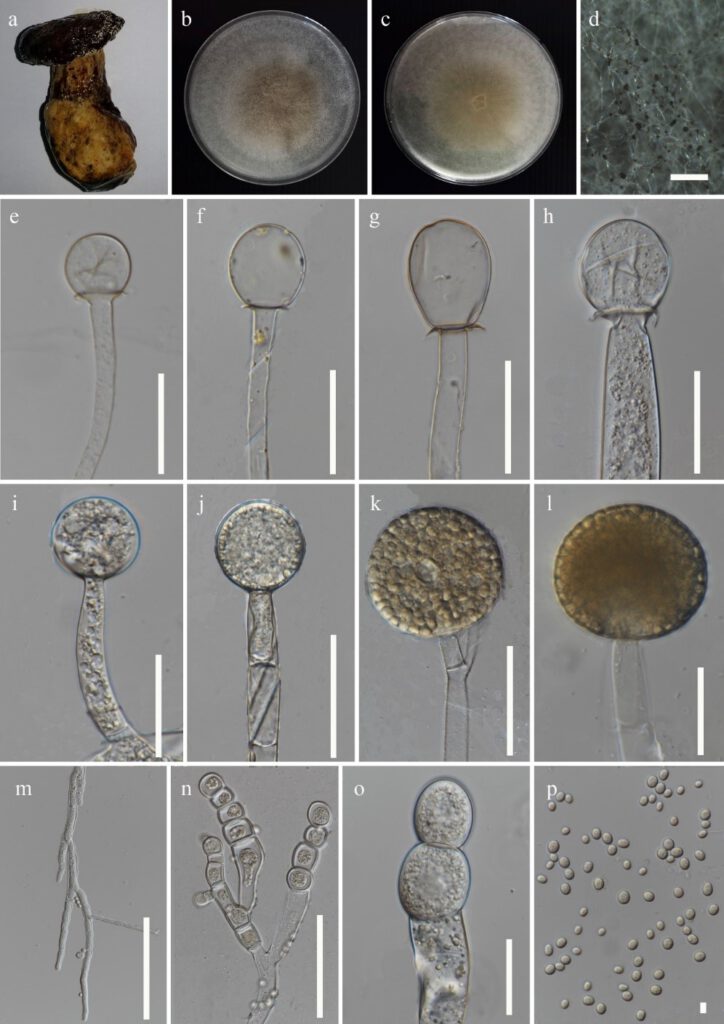
Mucor circinelloides Tiegh., Annls Sci. Nat., Bot., sér. 6 1: 94 (1875) Figure 3
MycoBank number: MB 198947; Index Fungorum number: IF 198947; Facesoffungi number: FoF 12292;
Fungicolous on a fruiting body of Phlebopus sp. Asexual morph on PDA: Sporangiophores up to 12–20 μm (x̅ = 15 μm, n = 20) in width, hyaline, repeatedly branched sympodially, having long and short branches, the latter infrequently circinate, short sporangiophores more profusely branched with short and often circinate branches, with slightly incrusted walls, the younger parts of the sporangiophores filled with granules, septate. Sporangia 40–53.5 × 39–53 μm (x̅ = 46.5 × 48.5 μm, n = 10), globose to sub-globose, initially hyaline and turning yellowish to become brownish-grey at maturity, decreasing in diameter in order of arisal on each sporangiophore, slightly incrusted walls; walls of the larger sporangia deliquescent and leaving small collars, and of the smaller sporangia persistent and leaving large basal membranes. Columellae 16–44 × 15–35 μm (x̅ = 30 × 25 μm, n = 20), obovoid to ellipsoidal in the larger sporangia, globose in the smaller sporangia, tinged a pallid brownish grey. Sporangiospores 4–7 × 3–6.2 μm (x̅ = 5.4 × 4.5 μm, n = 40) μm, ellipsoidal, smooth-walled, hyaline to brownish. Chlamydospores intercalary and terminal, variable in shape and size. Rhizoids hyaline. Sexual morph not observed.
Culture characteristics: on PDA, at 25 °C, cultures are initially white and later turn pale brown in the middle with sporangia formation. The colony reached 70 mm diam. after 4 days of incubation.
Materials examined: Thailand, Chiang Rai Province, Muang, Tha sut, isolated from the white mycelium growing on a fruiting body of Phlebopus sp., 25 June 2020, Gajanayake AJ, AJ 086 inactive dry culture MFLU xxxx, living culture MFLUCC xxxx.
GenBank accession numbers: ITS: xxx, LSU: xxx
Notes: Mucor circinelloides strain MFLU xxxx reported in this study shares similar asexual morphologies with those observed from the ex-neotype (CBS 195.68) of M. circinelloides described by Schipper (1976). During the micro-morphologcal examination, only 10 sporangia were measured as most of the sporangia were easily broken during the slide preparation due to the prevalence of deliquescent sporangial wall. The size of sporangia of MFLUCC xxxx (46.5 × 48.5 μm) is comparatively smaller than the sporangia of M. circinelloides (54 × 49 µm) described by Schipper (1976). The size of sporangiospores of MFLUCC xxxx (5.4 × 4.5 μm) is mostly similar to the sporangiospores of M. circinelloides (5.4 × 4 µm) described by Schipper (1976). The slight dimensional differences may have occurred due to the variations of agar medium and the conditions used to obtain the cultures. To the best of our knowledge, this is the first report of M. circinelloides on a fruiting body of Phlebopus sp. as a fungicolous fungus.

Figure 3: Mucor circinelloides (MFLU xxx) a Host b & c Colony on PDA d Sporulation on PDA e-h Columella with collars i-l Sporangia m Rhizoid n & o Chlamydospores p Sporangiospores. Scale bars: d = 200 μm, m = 150 μm, g,h,j,k,n = 50 μm, f,i,l,o = 30 μm, e = 25 μm, p = 5 μm.
