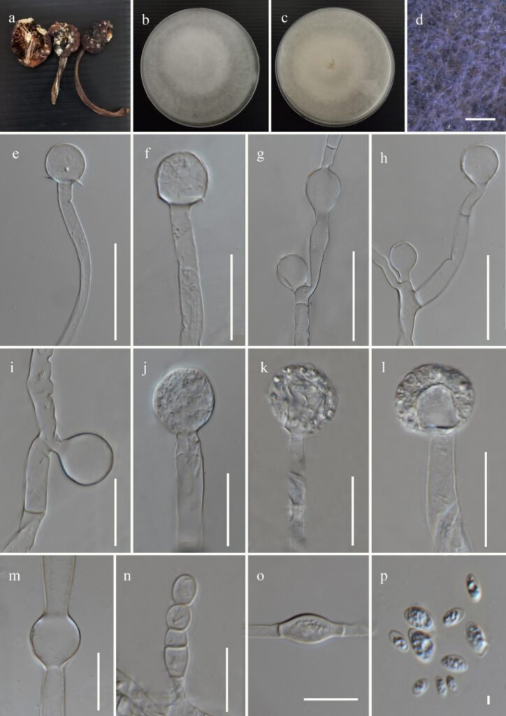
Mucor yunnanensis Gajanayake., Karun., Jayaward. & K.D Hyde, sp. nov. Figure 2
MycoBank number: MB; Index Fungorum number: IF; Facesoffungi number: FoF 12291;
Etymology: The epithet refers to the place “Yunnan Province” where the species was collected.
Holotype: ZHKU 22-0058
Fungicolous on a fruiting body of Armillara sp. Asexual morph on PDA: Sporangiophores up to 4.2–19.7 μm (x̅ = 9.6 μm, n = 50) width, hyaline, occasionally curved, septate, sympodial or monopodial branches formed. Sporangia 20–38 × 21.8–37 μm (x̅ = 29.6 × 30 μm, n = 12), globose, matured sporangial wall deliquescent, transparent and easily broken, hyaline. Columellae 13.5–31.5 × 10⁻³⁰ μm (x̅ = 21 × 19 μm, n = 35), mostly globose to sub globose, and rarely ellipsoid, collar present oftenly, hyaline to pale brown, smooth-walled. Sporangiospores 2.3–7.2 × 1.5–4.4 μm (x̅ = 4.7 × 2.5 μm, n = 50), mostly ellipsoidal, occasionally oval or irregular, smooth-walled, hyaline with granular content in the middle. Chlamydospores abundant, intercalary and terminal, variable in shape and size. Hyphal bulges were frequently observed. Sexual morph not observed.
Culture characteristics: on PDA, at 25 °C, cultures are cottony and white to pale yellow. The colony reached 60 mm diam. after 5 days of incubation.
Materials examined: China, Yunnan Province, Lijiang, Shigu, isolated from the white mycelium growing on a fruiting body of Armillara sp., 03 June 2020, Karunarathna SC, SG 069 Herbarium material ZHKU 22-0058 (Holotype), living ex-type culture ZHKUCC 22-0110; China, Yunnan Province, Lijiang, Shigu, isolated from the white mycelium growing on a fruiting body of Boletus sp., 29 June 2020, Karunarathna SC, SG 08B Herbarium material ZHKU 22-0057 (Paratype), living culture ZHKUCC 22-0109.
GenBank accession numbers: ZHKUCC 22-0110 (Ex-type)- ITS: xxx, LSU: xxx; ZHKUCC 22-0109 – ITS: xxx, LSU: xxx
Notes: Based on our phylogenetic analyses it was observed that, M. yunnanensis is phylogenetically distinct from M. irregularis, M. aseptatophorus and M. takensis and the placement was stable in both ML and BI analyses. Two strains of M. yunnanensis form a sister clade to the clade formed by M. irregularis, M. aseptatophorus, M. takensis with 97% bootstrap support. Simultaneously, M. yunnanensis is clustered within the sister clade formed to the clade consisting M. nidicola and M. merdicola. The synopsis of morphological characteristics related to the taxon introduced in this study and relevant sister taxa are shown in Table 3. During the micro-morphologcal examination of M. yunnanensis, only 12 sporangia were measured as most of the sporangia were easily broken during the slide preparation due to the prevalence of deliquescent sporangial wall. In contrast to M. irregularis, highly variable shapes in columellae and sporangiospores were not observed in M. yunnanensis as it forms mostly globose to sub globose, and rarely ellipsoid columellae and mostly ellipsoidal, occasionally oval or irregular sporangiospores. Furthermore, the sizes of sporangia, sporangiospores and columellae are higher in M. irregularis when compared with M. yunnanensis. In contrast to M. aseptatophorous, septa were observed below sporangia in M. yunnanensis. The size of sporangia is higher in M. aseptatophorous when compared with M. yunnanensis. In contrast to M. takensis, sporangiophores were septate in M. yunnanensis. The size of sporangiospores is smaller in M. takensis compared to M. yunnanensis while granular content is observed in sporangiospores of M. yunnanensis in contrast to M. takensis. Therefore, based on both phylogeny and morphology, M. yunnanensis is introduced as a new species.

Figure 2: Mucor yunnanensis (ZHKUCC 22-0110, holotype) a Host b & c Colony on PDA d Sporulation on PDA e-i Columella with and without collars j-l Immature and mature sporangia m Hyphal bulge n & o Chlamydospores p Sporangiospores. Scale bars: d = 100 μm, e-m = 30 μm, n = 20 o = 10 μm, p = 5 μm.
