Solomyces sinensis Zhi.Y. Zhang, Y.F. Han and Z.Q. Liang
MycoBank number: MB 835714; Index Fungorum number: IF 835714; Facesoffungi number: FoF 08687; (Figure 7).
Etymology: In reference to China, the country where the type specimen was obtained.
Holotype: permanently preserved in a metabolically inactive state, HMAS 255397.
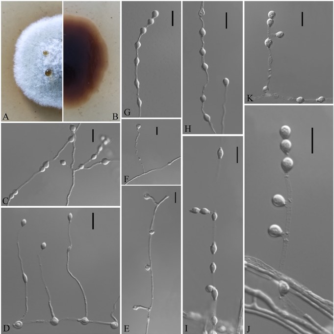
Description based on HMAS 255394. Asexual: Colonies on PDA, reaching 16–17 mm in diameter after 14 days at 25◦C, elevate at the center, velvety to floccose, margins regular, pewter at the center and white at the edge, producing a diffusible faint yellow pigment and clear exudates; reverse brown. Aerial mycelium abundant, smooth and thin-walled, septate, 1- to 2- µm wide. Terminal and lateral conidia born on hyphae, short protrusions, or side branches. Conidia solitary, sometimes two in chains, pyriform, sometimes subglobose, smooth or rarely rough walled, ecru, 4.0–6.0 3.0–5.5 µm (av. = 5.5 4.0, n = 50). Intercalary conidia abundant, olivary, subglobose to globose, 4.0–7.0 3.0–5.5 µm (av. = 5.0 4.0, n = 50). Sexual morph: not observed. Geographical distribution: China.
Material examined: China, Guizhou, Guiyang, the Affiliated Hospital of Guizhou Medical University, 26◦59rN, 106◦71rE, from soil beside a road, September 2016, Zhi. Y. Zhang (HMAS 255397 – holotype; CGMCC 3.18498 = GZUIFR K277.1 – ex-type living cultures; ibid., CGMCC 3.18499 = GZUIFR K277.2; ibid., CGMCC 3.18500 = GZUIFR K277.3). The living cultures were kept in sterile 30% glycerol and deposited in a 80 ◦C freezer.
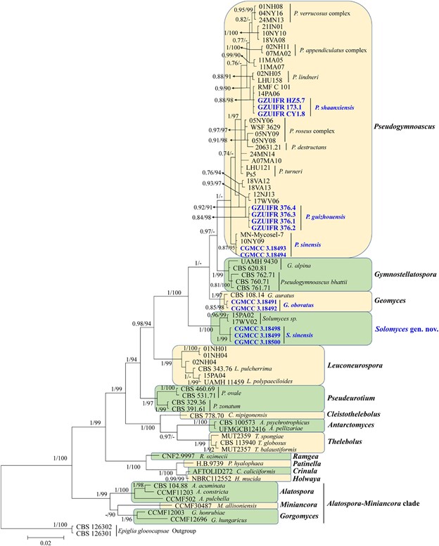
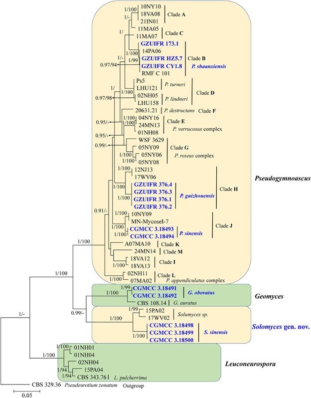
Notes: Solomyces sinensis was isolated from soil in Guizhou Province, China. We did not compare morphological characteristics between S. sinensis and another two strains within Solomyces owing to the lack of morphological description of these two strains (Minnis and Lindner, 2013). However, S. sinensis is phylogenetically distinct from these strains with high statistical support (1.00 BYPP, 100% MLBS) (Figures 1, 2).

FIGURE 1 | Phylogenetic relationships among the genera in Thelebolales based on Bayesian inference and maximum-likelihood analyses of the concatenated internal transcribed spacers (ITS) and LSU dataset. Posterior probabilities ≥0.7 and maximum-likelihood bootstrap values ≥90% are shown above the internal branches. The new isolates are presented in bold and blue.

FIGURE 2 | The consensus tree from the Bayesian inference and Geomyces and its allied genera based on the ITS, LSU, MCM7, RPB2, and EF1A concatenated dataset. Posterior probabilities ≥0.7 and maximum-likelihood bootstrap values ≥90% are shown above the internal branches. The new isolates are presented in bold and blue.

FIGURE 7 | Solomyces sinensis (from ex-holotype CGMCC 3.18498). (A,B) The front and reverse of a S. sinensis colony on PDA after 14 days at 25◦C. (C–K) Conidiophores and conidia. Scale bars = 10 µm.
