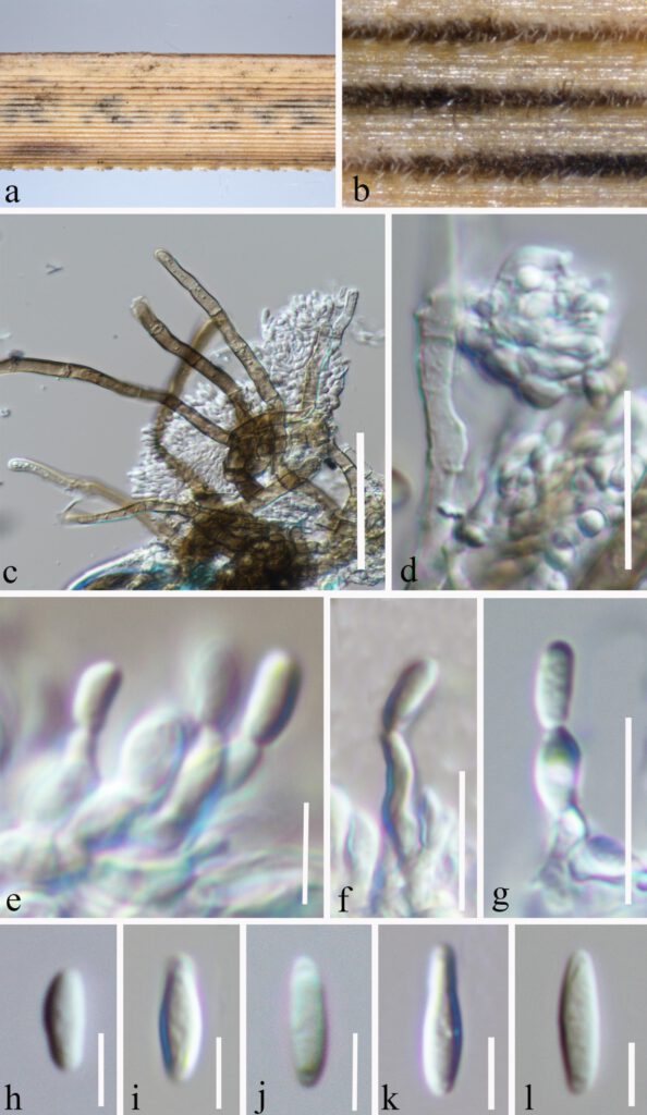
Microdochium kunmingense Y. Gao, H. Gui & K.D. Hyde, sp. nov.
MycoBank number: MB; Index Fungorum number: IF; Facesoffungi numbers: FoF 12705 ;
Etymology – Named after the location, Kunming from where the holotype was collected.
Holotype – HKAS123201
Saprobic on decaying herbaceous stem of grass. Sexual morph: Undetermined. Asexual morph: Hyphae 4–6.2 μm diam, (x̅ = 5.2 μm, n = 20), immersed and superficial, septate, hyaline or brown, unbranched or branched. Conidiogenous cells (4–)4.5–6(–6.4) × (1.7–)2–2.6(–3) μm (x̅ = 5 × 2 μm, SD = 0.7 × 0.3 μm, n = 15), phialidic, ampulliform, hyaline, smooth. Conidia (3.6–)5–9(–10.4) × (1.6–)2–2.8(–3) μm (x̅ = 7 × 2.4 μm, SD =2 × 0.4 μm, n = 20) fusiform or ellipsoidal to subcylindrical, aseptate.
Culture characteristics – Conidia germinated on PDA within 24 hours and germ tubes produced from one or both ends. Colonies on PDA reaching 30 mm in 4 weeks at room temperature (25–27°C), irregular, nmbonate, floccose, mycelia dense, pale pink from the above and center brown around light yellow from the below.
Material examined – CHINA, Yunnan Province, Kunming, Kunming Botanical Garden (25°8’19″N, 102°44’25″E), on decaying herbaceous stem of grass, 13 May 2021, Ying Gao (HKAS 123201, holotype), ex-type culture, CGMCC3.23526.
Known host and distribution – herbaceous stem of grass (China)
Note – Microdochium kunmingense grouped as a sister taxon to Microdochium rhopalostylidis with 99 % ML and 1.00 PP statistical support. Microdochium rhopalostylidis was introduced by Crous et al. (2019) and from the leaves of Rhopalostylis sapida (Arecaceae) in Auckland Botanical Garden, New Zealand. Morphologically, Microdochium rhopalostylidis has comparatively longer conidia (13–23 × 2.5–4 µm) to those of Microdochium kunmingense (3.6–10.4 × 1.6–3). Furthermore, the conidia of Microdochium kunmingense fusiform or ellipsoidal to subcylindrical, aseptate.But Microdochium rhopalostylidis fusoid 1–3-septate. Microdochium kunmingense differs from M. rhopalostylidis in 141/821 bp of rpb2 (17.17 %).

FIGURE 1| Microdochium kunmingense (HKAS 123201, holotype) on decaying leaves of grass. (a, b) Colonies on the host. (c, d) Vertical section of the colonies. (e–g) Conidiophores and Conidiogenous cells. (h–l) Conidia. Scale bars: (c) = 30 μm, (d) = 20 μm, (e) = 5 μm, (f,g) = 10 μm, (h–l) = 5 μm.
