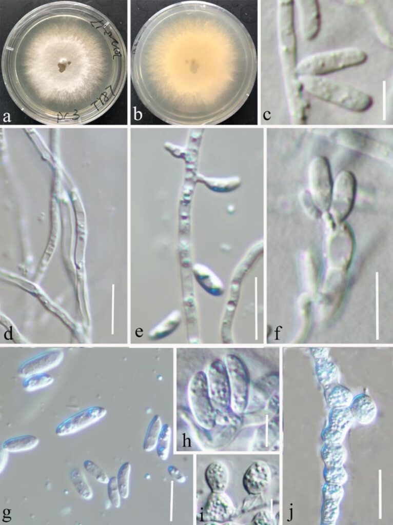
Microdochium setariae Y. Gao, H. Gui & K.D. Hyde, sp. nov. FIGURE 5.
MycoBank number: MB; Index Fungorum number: IF; Facesoffungi number: FoF 12706;
Etymology – The epithet refers to Setaria is the genus of the host plant (Setaria parviflora)
Holotype – HKAS 123195
Colonies on PDA attaining 56 mm in diameter after 15 days, flocculent, white to pale grey from above and reverse, becoming dark in the center after 4 weeks. Sexual morph: Undetermined. Asexual morph: Mycelium superficial, consisting of hyaline, finely verruculose, smooth, branched, septate, 1.5–3 µm wide hyphae. Chlamydospores 6–8.5 μm diam, thick-walled, subglobose or ovoid with constricted at medium, hyaline, granulate, terminal or intercalary, more frequently arranged in chains than clusters. Conidiogenous cells cylindrical or oblong shape, tapering towards both ends, hyaline, smooth, 0–1-septate, (12–)12.7–14.3(–14.6) × (3–)3.3–4(–4.3) μm (x̅ = 13.5 × 3.6 μm, SD = 0.8 × 0.3 μm, n = 15). Conidia aseptate, (6–)6.6–9(–10) × (2.3–)2.5–3.2(–3.8) μm (x̅ = 7.7 × 2.8 μm, SD = 1 × 3 μm, n = 30), subcylindrical, ellipsoid or lunate, aseptate, hyaline, smooth-walled, straight or curved with obtuse apex.
Known host and distribution – Setaria parviflora (China)
Culture characteristics – Colonies on PDA 50–60 mm in diameter after 15 days at room temperature, mycelia circular, flat, dense, the edges are failamcntous and white, grey at center, aerial mycelia cottony or sparse, reverse white.
Material examined – China, Yunnan Province, Kunming, Kunming Botanical Garden (25°8’19″N, 102°44’25″E), on healthy leaves of Setaria parviflora, 8 March 2021, Ying Gao (HKAS 123195, holotype), ex-type culture CGMCC3.23528; ibid., on leaves of healthy Setaria parviflora, 8 March 2021, Ying Gao (HKAS 123194 paratype), ex-paratype culture CGMCC3.23527; HKAS 123196, living culture CGMCC3.23529; HKAS 123197, living culture CGMCC3.23530.
Notes – Phylogeny of a concatenated LSU-ITS-tub2-rpb2 sequence dataset depicts Microdochium setariae as a sister taxon of M. bolleyi with 100 % ML and 1.00 PP statistical support (Fig. 1). Microdochium setariae resembles M. bolleyi in having hyaline, smooth conidiogenous cells, aseptate, hyaline, ellipsoid conidia (Hoog & Hermanides 1977). However, Microdochium setariae differs from M. bolleyi in having cylindrical conidiogenous cells (12–14.6 × 3–4.3 μm) and larger conidia (6–10 × 2.3–3.8 vs. 5.5–8.5 × 1.6–2.2 μm), while M. bolleyi has globose or subglobose conidiogenous cells (2–4.5 × 2–3.5 μm) (Hoog & Hermanides 1977, Table 5). A nucleotide pairwise comparison showed that Microdochium setariae differs from M. bolleyi in 16/821 bp of rpb2 (1.95 %, without gaps), 9/765 bp of tub2 (1.18 %).

FIGURE 1| Microdochium setariae (HKAS 123195, holotype). On leaves of healthy Setaria parviflora. (a) Surface of colony on PDA. (b) Reverse of colony on PDA. (d) Hyaline mycelium. (c, e, f) Conidiophores and conidiogenous cells. (g, h) Conidia. (i, j) Chlamydospores. Scale bars: (c) = 5 μm, (d–g) = 10 μm, (h,i) = 5 μm, (j) = 15 μm.
