Microdochium shilinense Y. Gao, H. Gui & K.D. Hyde, sp. nov.
MycoBank number: MB; Index Fungorum number: IF; Facesoffungi number: FoF 12704;
Etymology – Named after the location (Shilin County), from where the holotype was collected.
Holotype – HKAS 123198
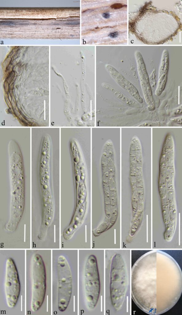
Saprobic on dead herbaceous stem of grass. Sexual morph: Ascomata 125–150 μm diam × 100–120 μm high, (x̅ = 134 × 111 μm, n = 10), scattered, gregarious, deeply immersed in host tissues, globular or subglobose, light brown to black, uni-loculate, without ostiolate, slightly raised top. Peridium 10–20 μm thick, (x̅ = 12 μm, n = 30), composed of 3–4 layers of flattened, thick-walled, light brown to dark brown cells of textura angularis. Paraphyses 3–4.5 μm wide, (x̅ = 3.7 μm, n = 20), straight or curved, septate, hyaline, unbranched, with large to small guttules, slightly constricted at the septa, filiform to strip. Asci (50–)52–67(–76) × (7–)8–9.6(–10) μm (x̅ = 60 × 8.8 μm, SD = 7 × 1 μm, n = 20), 8-spored, arising from base, cylindrical, bitunicate, with a short pedicel, hyaline, with refractive ring around cytoplasmic protrusion. Ascospores (14–)15–17(–18) × (3–)3.7–4.8(–5.7) μm (x̅ = 16 × 4.2 μm, SD = 1 × 0.5 μm, n = 30), overlapping, 2-seriate, hyaline, guttulate, fusiform, straight or curved, with 0–3 transverse septa, sometimes slightly constricted at medium septum, rounded to slightly pointed at both ends. Asexual morph: Undetermined.
Culture characteristics – Ascospores germinated on PDA within 24 hours at room temperature. Germ tube initially produced from the middle cell of ascospore. Colonies on PDA reaching 50 mm diameter after 4 weeks at 25–27°C, circular, slightly raised, smooth, fimbriate, filiform, floccose, white from the above and yellowish from the below.
Material examined – China, Yunnan Province, Kunming, Shilin County (24°49’23″N, 103°32’11″E), on decaying herbaceous stem of grass, 13 June 2021, Ying Gao (HKAS 123198, holotype), ex-type culture CGMCC3.23531.
Known host and distribution – erbaceous stem of grass (China)
Notes – The present phylogenetic analysis showed that Microdochium shilinense forms a distinct branch as the basal clade of M. seminicola, M. graminearum and M. albescens with high bootstrap support (100% ML-BS and 1.00 BYPP, Fig. 1). The nucleotide pairwise comparison showed that M. shilinense differs from M. albescens in 46/765 bp of tub2 (6.01%) and 20/522 bp of ITS (3.83%). Microdochium shilinense differs from Microdochium seminicola and M. graminearum in having cylindrical asci with refractive ring around cytoplasmic protrusion, while M. seminicola has fusiform asci with funnel-shaped apical ring; M. graminearum has fusiform and comparatively larger asci (55–77.6 × 9.6–15.5 μm). Microdochium shilinense differs from Microdochium albescens in having fusiform ascospores with 0–3 transverse septa, while M. albescens has fusoid ascospores with >3 transverse septa.

FIGURE 1. Microdochium shilinense (HKAS 123198, holotype) (a, b) Appearance of immersed ascomata on the host. (c) Vertical section of the ascoma. (d) Peridium. (e) Paraphyses. (f–l) Asci. (m–q) Ascospores. (r) Culture characters on PDA. Scale bars: (c–f ) = 20 μm, (g–j) = 10 μm, (k,l) = 20 μm, (m–q) = 10 μm.
