Melanconiella flavovirens (G.H. Otth) Voglmayr & Jaklitsch, in Voglmayr, Rossman, Castlebury & Jaklitsch, (2012)
MycoBank number: MB 800120; Index Fungorum number: IF 800120; Facesoffungi number: FoF 11775; Fig. 23
≡ Diaporthe flavovirens G.H. Otth, (1869)
= Melanconis flavovirens (G.H. Otth) Wehm., (1937) (see Index Fungorum 2022, and Voglmayr et al. 2012)
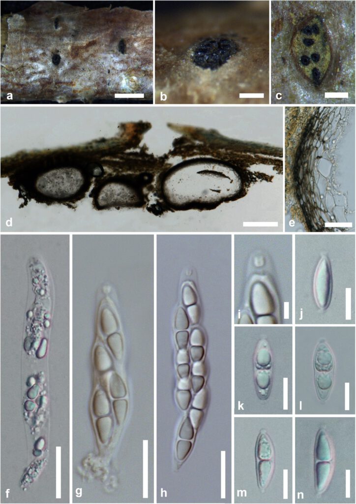
Saprobic on dead aerial branch of Corylus avellana L. Sexual morph: Pseudostromata 1.2–2.5 mm diam. (x̅ = 1 × 2 mm, n = 10), scattered on substrate, circular or elliptical, erumpent, projecting up to 200–400 μm, arranged with perithecial bumps, view as raised black dots. Ectostromatic disc 0.4–1.2 mm diam, elliptic or circular outline, pulvinate, grayish-yellow. Entostroma more or less well developed, yellowish to pale brown. Ostioles 8–10 per disc, unevenly emerging on the disc, circular, slightly papillate, black. Perithecia 300–600 mm wide, immersed in host bark, confluent. Peridium 15–20 μm wide, 15–25 μm wide at base, composed of 5–7 layers, outermost layers comprising dark brown to pale brown cells of textura prismatica, fused with host tissues, inner layer comprising pale brown to hyaline cells of textura angularis. Asci 60–100 × 13–16 (x̅ = 85 × 13.5 μm, n = 20) μm, 8-spored, untunicate, sessile and rounded pedicel, distinct apical ring with 2–3 μm wide. Ascospores 18–22 × 5–7 μm (x̅ = 20 × 6.5 μm, n = 40), uni-biseriate, overlapping, ellipsoid or broadly fusoid, rounded or subacute at the apices, 1-septate, not constricted at the septum, hyaline, with distinct and persistent knob-like appendages with 1–2 μm long, commonly monomorphic upper and lower cells, cells distinctly triangular-ovate in outline, with two large or numerous small guttules. Asexual morph: discosporina-like. see detailed description in Voglmayr et al. (2012).
Material examined – Italy, province of Forlì-Cesena [FC], Massera – Predappio, dead and hanging branch of Corylus avellana L. (Betulaceae), 10 February 2019, Erio Camporesi, IT 4222, (MFLU 19-0641), ibid., 15 February 2019, IT 4222a (MFLU 19-0642).
GenBank numbers – ITS: OM614593; LSU: OM616564; rpb2: ***, tef1-α:***.
Notes – Myxosporium sulphureum (asexual morph) was described by Saccardo (1884) based on the description provided by Fuckel (1871). The specimen from the Fuckel herbarium for Myxosporium sulphureum was designated as the lectotype of Melanconiella flavovirens by Voglmayr et al. (2012) based on morphological similarities. Our strain (MFLU 19-0641) is morphologically similar to the type collection of Melanconiella flavovirens (CBS 125598, MFV3, MFV1), in having large ectostromatic discs, triangular to ovate cells and knob-like appendages of ascospores (Voglmayr et al. 2012, this study). Asexual morph is less prominent and unavailable from fresh cultures, while a single collection of conidiomata was collected from Italy (Voglmayr et al. 2012). Phylogenetic analysis placed our strains MFLU 19-0641 with Melanconiella flavovirens isolates (CBS 125598, MFV3, MFV1) with 100% MLBS, 1.00 BYPP support (Fig. 25). Therefore, we identified our new collection as Melanconiella flavovirens from Corylus avellana (Betulaceae) in Italy.
The majority of phylogenetically distinct sexual taxa of Melanconiella could also be identified based on morphology (Voglmayr et al. 2012). Some sexual morph characters are not identical with the morphology of phylogenetically closely related taxa. Therefore, host association and sexual-asexual linkages may help to identify the taxa (Voglmayr et al. 2012). The majority of Melanconiella species are highly host-specific and recorded on Fagalae trees, such as Betula sp., Betula pendula, Carpinus betulus, C. caroliniana, C. orientalis, Corylus avellana, Ostrya carpinifolia, and O. virginiana in Europe (Voglmayr et al. 2012). Melanconiella flavovirens was first reported on the Corylus avellana tree from the Lombardia region in northern Italy (Voglmayr et al. 2012), and our new collection is the first record from Emilia–Romagna in northern Italy.

Figure 23 – Melanconiella flavovirens (MFLU 19-0641). a–c. Pseudostromata on a dead branch of Corylus avellana. c, d. Transverse and longitudinal sections of pseudostromata. e. Peridium. f–h. Asci. i. well distinct apical ring. j–n. Ascospores. Scale bars: a = 1 mm, b, c = 400 µm, d = 200 µm, e–h= 20 µm, j–n= 10 µm, i = 5 µm.
