Description
Pileus 10-16 mm diam., convex to umbonate, expanding to plano-concave, with straight margin; rough or with crowded squamules at center, light brown to brown (7D6-8, 7E7-8) at center, with concolourous squamules around umbo towards margin, slightly distant at marginal zone on white to orange white (5A2) blackground; margin broken, sulcate or striate, appendicular, with concolourous squamules on surface and white fibrillose remnants. Lamellae free, broadly ventricose, 3-4 mm wide, white, moderately crowded, with 1 length lamellulae, with eroded edge. Stipe 25-30 × 3-4 mm, cylindrical, slightly wider at base; completely fibrillose, crowded at annular zone down toward base, white, with light brown to brown (7D6-8, 7E7-8) squamules at base zone, on white to orange-white (5A2) background. Annulus with an annular zone or cortinate, white. Context white in pileus, up to 1 mm wide; hollow in stipe, concolorous with surface. Taste and smell unknown. Spore print white.
Basidiospores [50,2,2] 5.0-6.0 × 3.2-4.0 µm, avl × avw=5.54 × 3.64 µm, Q= 1.45-1.57, Qav= 1.52, in side-view ellipsoid ovoid, in frontal view ovoid to, slightly thick-walled, dextrinoid, congophilous, cyanophilous, not metachromatic. Basidia 15.0-18.0 × 5.0-7.0 µm, clavate, slightly thick-walled, hyaline, 4-spored. Cheilocystidia 18.5-29.0 × 5.0-10.0 µm, narrowly clavate to clavate, rarely broadly clavate, slightly thick-walled, hayaline. Pleurocystidia absent. Pileus covering a trichoderm made up of two layer of elements; upper layer made up of cylindrical to narrowly cylindrical elements with rounded apex, sometimes swollen at middle and tapering to base and apex, 60-140 × 9.0-17.5 µm, with pale brown pale brown parietal and intracellular pigment; underlayer made up of shortly clavate elements, 30.0-45.0 × 10.0-20.0 µm, with pale brown pale brown parietal and intracellular pigment. Stipe covering of squamules at base zone similar to pileus covering. Clamp connections present in all tissues.
Habitat and distribution: solitary, saprotrophic, on decayed humus soil; found in high elevation deciduous forests of northern Thailand.
Material examined: Thailand, Chiang Mai Province: Mae Taeng district, Pha Deng village, N 19° 07.13′, E 98° 43.52′, 905 m, 04 July 2010, P. Sysouphanthong P98 (MFLU 09-0616, holotype); ibidem, 25 July 2008, P. Sysouphanthong PS093 (MFLU 09-0166, paratype).
Note: Lepiota subthailandica has a tiny basidioma, a trichodermal structure of pileus covering and ellipsoid ovoid basidiospores; and the species is located in Lepiota sect. Ovidsporae (J.E. Lange) Kühner (Velliga 2001). In the same section, Lepiota subthailandica is very similar to L. thailandica Sysouph., K.D. Hyde, J.C. Xu & P.E. by morphology; and they are widespread in the same location of Chiang Mai, northern Thailand. However, L. thailandica has smaller basidiomata (3-4 mm diam. in pileus), utriform and fusiform cheilocystidia, and shorter elements of pileus covering (55–95 × 5.5–22 µm) (Sysouphanthong et al. 2016). Second species, L. microcarpa Sysouph., K.D. Hyde & Vellinga, is similar in micromorphology. However, L. microcarpa has penguin-shaped basidiospores, and belongs to sect. Lepiota (Sysouphanthong et al. 2012). Based on the nrITS sequences analysis, two samples of L. subthailandica are not related to L. thailandica, and separated from other species in the sect. Ovisporae.
GenBank numbers:
ITS: MT436254, MT436253 (ITS1-F/ITS4).
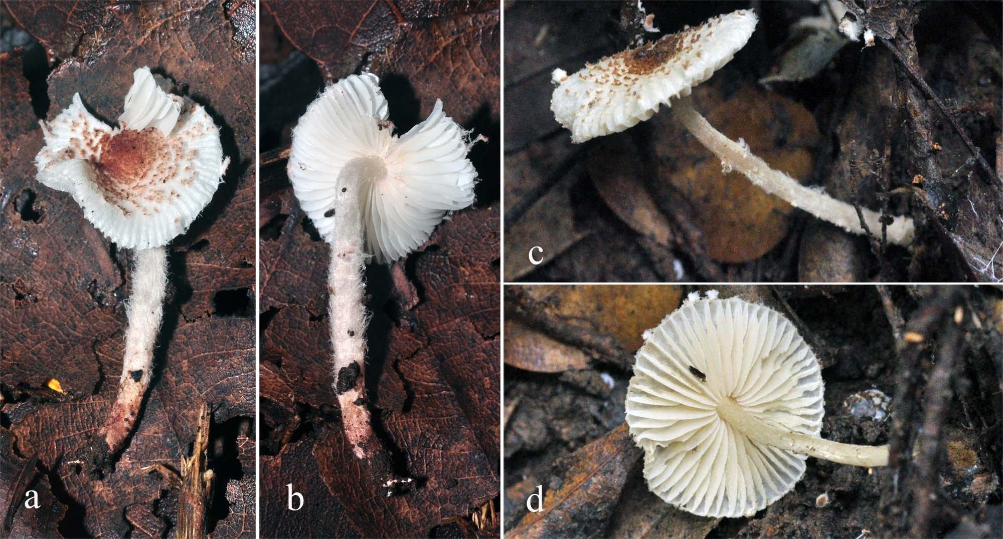
Figure 1. Lepiota subthailandica in habitat. a-b= MFLU 09-0166; c-d= MFLU 10-0616, holotype).

Figure 2. Lepiota subthailandica (holotype, MFLU 10- 0616), a= basidiospores, b= basidia, c= cheilocystidia, d= pileus covering.