Description
Pileus 30-65 mm, sub umbonate to umbonate, expanding to plano-concave, with inflexed margin; greyish orange to brownish orange (6B5-8, 6C7-8) glabrous at umbo, with concolorous squamules toward margin on white fibrillose background; margin sulcate, with partial veil remnants, concolorous with squamules on surface. Lamellae free, broadly ventricose, 3-5 mm wide, white, moderately crowded, with 3 length lamellulae, with eroded edge. Stipe 60-80 × 5-4 mm, cylindrical or slightly tapering to apex, completely fibrillose or cortinate at annular zone, white, with white to greyish orange to brownish orange (6B5-8, 6C7-8) squamules under annular zone downward base, on white to orange-white (5A2) background. Annulus an annular zone, cortinate or fibrillose, white. Context in pileus white, up to 4 mm wide; in stipe hollow, concolorous with surface. Smell and taste unknown. Spore print white.
Basidiospores [50,2,2] 13.0-16.5 × 4.0-5.0 µm, avl × avw=17.7 × 4.7 µm, Q= 2.91-3.30, Qav= 3.20, in side-view cylindrical, with attenuate or rounded apex, with straight abaxial side, with inflexed hilar appendage, with suprahilar depression, fusiform to cylindrical in frontal view, slightly thick-walled, hyaline, dextrinoid, congophilous. Basidia 22-33 × 7.0-9.0 µm, clavate, slightly thick-walled, hyaline, 4-spored. Cheilocystidia 15-35 × 5.0-20.0 µm, clavate to broadly clavate, sometimes utriform, branched or with septates under element cell, thick-walled, hyaline. Pileus covering a trichoderm made up of two layers of elements; upper layers made up of cylindrical elements with rounded or attenuate apex, 70-190 × 5.0-13.0 µm, hyaline to pale brown, slightly thick-walled, smooth-walled, with parietal pigment; under layers made up of shortly clavate to clavate elements, 35-50 × 8.0-14.0 µm, smooth or rough-walled, with hyaline to parietal pale brown pigment. Stipe covering of squamules similar to pileus covering. Clamp connections present.
Material examined: Laos, Oudomxay Province, Xay District, Houay Houm Village, N 20° 32′ 00.67″, E 101° 53′ 48. 17.16″. 917 m., 15 June 2014, P. Sysouphanthong, PS2014-1465 (HNL503136); ibidem, 20 July 2014, P. Sysouphanthong, PS2014-1480 (HNL503151).
Habitat and distribution: solitary or grow in a small cruster with few basidiomycota, on dead leaves and humus soil, saprotrophic.
Notes: Lepiota metulispora has a trichodermal pileus covering and penguin-shaped basidiospores, and it is placed in sect Lepiota (Vellinga 2001). The species is widespread in tropical countries. Lao specimens were found in mature stage, colour of squemules on pileus is paler than Thai specimens, but the morphology is totally identical (Sysouphanthong et al. 2012). The type specimens of the species from Sri Lanka closes to Lao and Thai specimens by morphology, but basidiospores are slightly lager and cheilocystidia are unknown (Peler, 1972). Liang et al. (2011) studied the type material from Sri Lanka and compared with Chinese specimens, and the type specimens has smaller basidiospore size. However, it seems that size of basidiospores are not much different in all specimens found in China, Laos, Sri Lanka and Thailand; and the morphology and size can be minorly different in different specimens.
GenBank numbers:
ITS: MT436261, MT436262 (ITS1-F/ITS4).
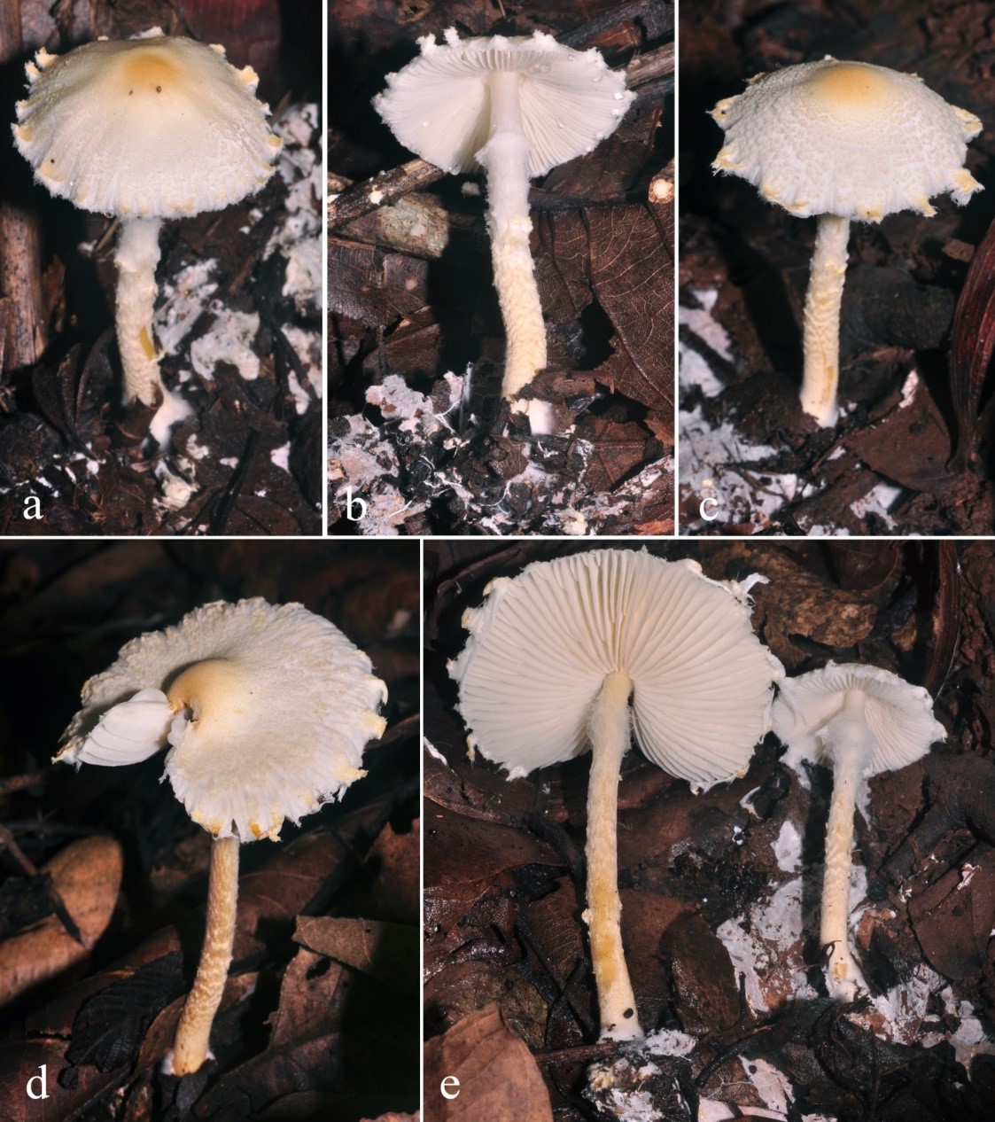
Figure 1. Lepiota metulispora in habitat, a-c= HNL503136, d-e= HNL503151.

Figure 2. Lepiota metulispora (HNL503151). a= basidiospores, b= cheilocystidia, c= pileus covering.