Lasiodiplodia microconidia Y. Zhang ter & S. Lin, Mycol. Progr. 18(5): 693 (2019)
MycoBank number: MB 831469; Index Fungorum number: IF 831469; Facesoffungi number: FoF 10664; Fig. **
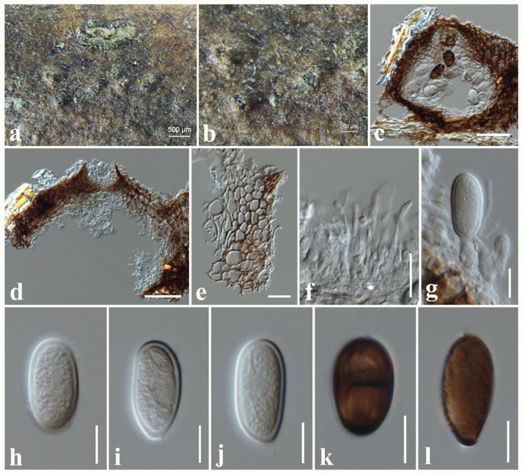
Saprobic on dead twigs attached to Anomianthus dulcis. Sexual morph: Undetermined. Asexual morph: Coelomycetous. Conidiomata 150–180 μm high × 150–230 μm diam. ( = 160 × 180 µm, n = 10), pycnidial, dark brown, globose to subglobose, solitary, scattered, immersed to semi-immersed, uni-locular, with a central ostiole. Conidiomata wall 20–30 μm wide, composed of brown cells of textura angularis. Paraphyses up to 40 μm long, 1–2 μm wide, hyaline, cylindrical, septate, rounded at apex. Conidiophores reduced to conidiogenous cells. Conidiogenous cells 8–12 × 4–6 μm ( = 10 × 5 µm, n = 10), hyaline, holoblastic, discrete, cylindrical to subcylindrical, smooth-walled. Conidia 20–30 × 12–15 μm ( = 26 × 13 µm, n = 30), initially hyaline, subglobose to subcylindrical, with granular content, both ends rounded, wall <2 μm thick, becoming pigmented, ellipsoid to ovoid, 1-septate with longitudinal striations.
Culture characteristics – Colonies on PDA reaching 80 mm diameter after 2 days at 25°C, colonies from above: olivaceous-grey, circular, margin entire, fluffy appearance with abundant aerial mycelia; reverse: light brown.
Material examined – THAILAND, Chiang Rai Province, dead twigs attached to Anomianthus dulcis (Annonaceae), 4 April 2019, N. I. de Silva, AND1 (MFLU 21-0226), living culture, MFLUCC 21-0191.
Known hosts and distribution – From healthy tissues of Aquilaria crassna in Laos (Wang et al 2019).
GenBank numbers – ITS: *******, tef1: ********, tub2: *******.
Notes – Lasiodiplodia microconidia was introduced by Wang et al (2019) from healthy tissues of Aquilaria crassna in Laos. Phylogeny indicates that our strain clusters with the type L. microconidia (Fig. ***). Conidia of new collection (26 × 13 µm) are slightly larger than the type (20.8 × 13.2 μm). This is the first record of L. microconidia from dead twigs of Anomianthus dulcis.

Figure ** – Lasiodiplodia microconidia (MFLU 21-0226). a, b Appearance of conidiomata on the substrate. c, d Sections through conidioma. e Peridium. f Paraphyses. g Conidiogenous cells. h–l Conidia. Scale bars: a = 500 μm, b = 200 μm, c, d = 50 μm, e, f = 20 μm, g–l = 10 μm.
