Lasiodiplodia aquilariae Y. Zhang ter & S. Lin, Mycol. Progr. 18(5): 685 (2019)
MycoBank number: MB 821001; Index Fungorum number: IF 821001; Facesoffungi number: FoF 10663; Fig. **
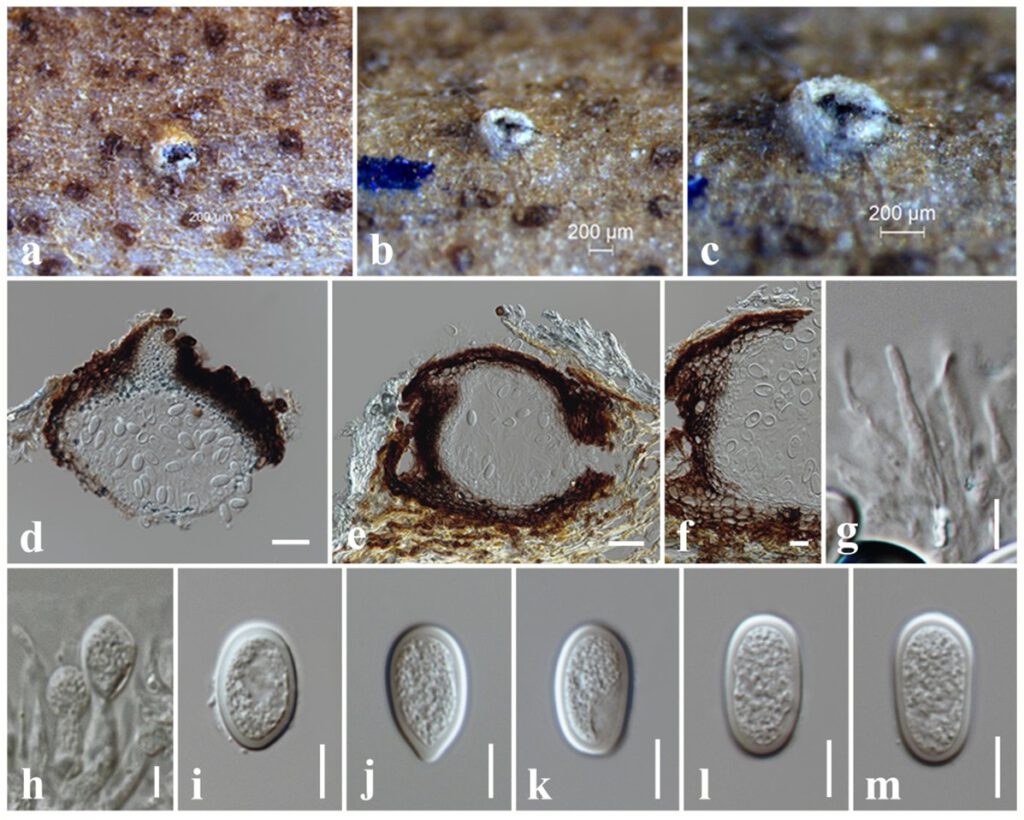
Saprobic on dead twigs attached to Magnolia champaca. Sexual morph: Undetermined. Asexual morph: Coelomycetous. Conidiomata 250–260 μm high × 270–295 μm diam. ( = 255 × 284 µm, n = 10), pycnidial, brown, globose to subglobose, solitary, immersed to semi-immersed, erumpent through plant host tissue. Conidiomata wall 30–40 μm wide, composed of brown cells of textura angularis. Paraphyses up to 55 μm long, 2–3 μm wide, hyaline, cylindrical, septate, rounded at apex. Conidiophores reduced to conidiogenous cells. Conidiogenous cells 7–13 × 3–5 μm ( = 11 × 4 µm, n = 10), hyaline, holoblastic, discrete, cylindrical to subcylindrical, smooth-walled. Conidia 20–27 × 10–13 μm ( = 25 × 12 µm, n = 30), hyaline, subglobose to subcylindrical, with granular content, both ends rounded, wall <2 μm thick.
Culture characteristics – Colonies on PDA reaching 50 mm diameter after 3 days at 25°C, colonies from above: white, circular, margin entire, dense, cottony to fluffy appearance with abundant aerial mycelia; reverse: cream.
Material examined – THAILAND, Chiang Rai Province, dead twigs attached to Magnolia champaca (Magnoliaceae), 9 January 2019, N. I. de Silva, NI305 (MFLU 21-0224), living culture, MFLUCC 21-0184.
Known hosts and distribution – From Aquilaria crassna in Laos (Wang et al. 2019).
GenBank numbers – ITS: *******, tef1: ********, tub2: *******.
Notes – Morphologically, a new collection (MFLU 21-0224) resembles L. aquilariae in having similar size range of conidia. Conidia of the new isolate are (20–27 × 10–13) μm and L. aquilariae are (25–28 (–29) × 12–16) μm (Wang et al. 2019). Phylogeny based on a combined ITS, tef1 and tub2 sequence data revealed the new isolate closely related to L. aquilariae with 54% ML and **** BYPP statistical support (Fig. **). A pairwise comparison of tef1 sequence data between the new isolate MFLUCC 21-0184 and L. aquilariae shows four base pair (1.34%) differences. ITS gene region of both the new isolate and L. aquilariae are similar. A comparison of tub2 sequence data was not done as it is not available for L. aquilariae in GenBank. Lasiodiplodia aquilariae was described in Laos from Aquilaria crassna (Wang et al. 2019). This is the first report of L. aquilariae from dead twigs of Magnolia champaca in Thailand.

Figure ** – Lasiodiplodia aquilariae (MFLU 21-0224). a–c Appearance of conidiomata on the substrate. d, e Sections through conidioma. f Peridium. g Paraphyses. h Conidiogenous cells. i–m Conidia. Scale bars: b, c = 200 μm, d, e = 50 μm, f = 20 μm, g–m = 10 μm.
