Lasiodiplodia pyriformis F.J.J. Van der Walt, Slippers & G.J. Marais, Persoonia 33: 163 (2014)
MycoBank number: MB 518722; Index Fungorum number: IF 518722; Facesoffungi number: FoF 10665; Fig. **
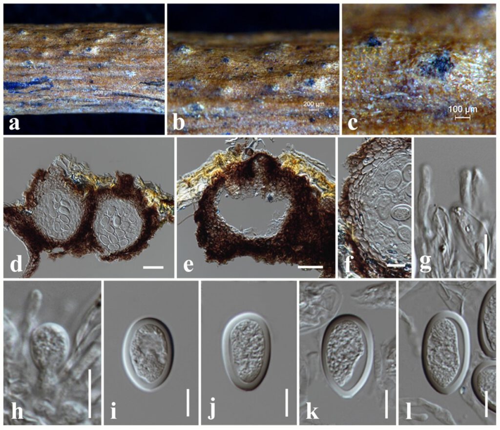
Saprobic on dead twigs attached to Magnolia lilifera. Sexual morph: Undetermined. Asexual morph: Coelomycetous. Conidiomata 190–220 μm high × 180–230 μm diam. ( = 200 × 215 µm, n = 10), pycnidial, dark brown, globose to subglobose, solitary to gregarious, immersed to semi-immersed, erumpent through plant host tissue. Conidiomata wall 25–35 μm wide, composed of light brown cells of textura angularis. Paraphyses up to 30 μm long, 3–4 μm wide, hyaline, cylindrical, septate, rounded at apex. Conidiophores reduced to conidiogenous cells. Conidiogenous cells 6–10 × 5–6 μm ( = 7 × 5.3 µm, n = 10), hyaline, holoblastic, discrete, cylindrical to subcylindrical, smooth-walled. Conidia 24–30 × 16–18 μm ( = 26 × 17 µm, n = 30), hyaline, subglobose to subcylindrical, with granular content, both ends rounded, wall <2 μm thick.
Culture characteristics – Colonies on PDA reaching 55 mm diameter after 5 days at 25°C, colonies from above: light grey, circular, margin entire, slightly dense, cottony to fluffy appearance with abundant aerial mycelia; reverse: cream.
Material examined – THAILAND, Chiang Rai Province, dead twigs attached to the Magnolia lilifera (Magnoliaceae), 11 February 2019, N. I. de Silva, NI326 (MFLU 21-0230), living culture, MFLUCC 21-0190.
Known hosts and distribution – From the leading edges of lesions on branches of Senegalia mellifera in Nambia (Slippers et al 2014).
GenBank numbers – ITS: *******, tef1: ********, tub2: *******.
Notes – We have collected a fungal species from dead twigs of Magnolia lilifera and identified as Lasiodiplodia pyriformis based on the phylogeny of combined ITS, tef1 and tub2 sequence data (Fig. **). Conidia of the new isolate (26 × 17 µm) are longer than the type of L. pyriformis (23.3 × 17.6 µm) (Slippers et al 2014). Conidiogenous cells of the new isolate (7 × 5.3 µm) are smaller than the type of L. pyriformis (12.3 × 4.7µm) (Slippers et al 2014). Comparisons of DNA sequences between the new isolate and the ex-type L. pyriformis CBS 121770 revealed two base pairs (0.42%) and one base pairs (0.39%) differences in ITS and tef1 gene regions respectively. DNA sequences of tub2 gene region of both the new isolate and the ex-type L. pyriformis CBS 121770 are similar. Lasiodiplodia pyriformis was introduced by Slippers et al (2014) from lesions on branches of Senegalia mellifera in Nambia. This is the first record of L. pyriformis from dead twigs attached to the host plant of Magnolia lilifera in Thailand.

Figure ** – Lasiodiplodia pyriformis (MFLU 21-0230). a The specimen. b, c Appearance of conidiomata on the substrate. d, e Sections through conidioma. f Peridium. g Paraphyses. h Conidiogenous cells. i–l Conidia. Scale bars: b = 200 μm, c = 100 μm, d, e = 50 μm, f = 20 μm, g–l = 10 μm.
