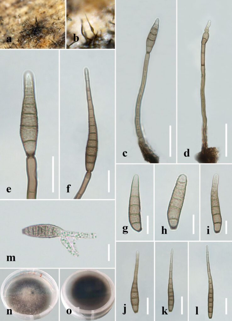
Distoseptispora aquamyces R. Zhu & H. Zhang, sp. nov.
MycoBank number: MB; Index Fungorum number: IF; Facesoffungi number: FoF 12576;
Description (Description with measurements of sexual and asexual state of species)
Saprobic on decaying wood submerged in freshwater. Sexual morph: undetermined. Asexual morph: hyphomycetous. Colonies on the substratum: superficial, effuse, scattered, hairy, brown to dark-brown. Mycelium partly superficial, partly immersed, composed of branched, septate, smooth, hyaline to pale-brown hyphae. Conidiophores (78–)91–198 μm long (x̄=127 µm, n=10), 4–7 μm wide (x̄=5 µm, n=10), macronematous, mononematous, unbranched, multi-septate, single or in groups of two or three, cylindrical, straight or slightly flexuous, smooth, pale-brown. Conidiogenous cells 10–20 μm long (x̄=16 µm, n=10), 4.5–5.5 μm wide (x̄=5 µm, n=10), monoblastic, integrated, determinate, terminal, cylindrical, pale-brown, smooth. Conidia 30–95 μm long (x̄=73 µm, n=20) (rostrum included), 7–12 µm at the widest part (x̄=8 µm, n=20), 2–5 µm wide at the apex (x̄=3 µm, n=20), acrogenous, dry, obclavate to obpyriform, mostly rostrate, straight or curved, 4–10-euseptate, mostly 6–9-euseptate, tapering towards the rounded apex, verrucose, thin-walled, pale-brown to brown at the base, subhyaline to pale-brown at the apex, verrucose.
Material examined: China, Sichuan Province, Yibin City, Southern Sichuan Bamboo Sea, Qicai Lake, found on dead, submerged, decaying wood of unidentified plants, 3 November 2020, Yun Qing, SN–18 (HKAS 122186, holotype), ex-type living culture KUNCC 21–10731.
Distribution: China
Sequence data: ITS: OK341187 (ITS5/ITS4); LSU: OK341199 (LROR/LR5); TEF1a: OP413482 (983/2218R); RPB2: OP413476 (fRPB2-5F/fRPB2-7cR)
Notes: Distoseptispora aquamyces differs from D. suoluoensis in having pale-brown to brown, shorter conidia (30–95 μm vs. 80–125 μm), sometimes with percurrent proliferation, while D. aquamyces has yellowish-brown or dark-olivaceous conidia without prolif-eration. In our phylogenetic tree, Distoseptispora aquamyces is close to D. suoluoensis, with low support. Two species in Distoseptispora possess verrucose conidia, i.e., D. suoluoensis and D. verrucose. Distoseptispora verrucose has percurrently proliferating conidiogenous cells and olivaceous conidia which differ from the non-proliferating conidiogenous cells and pale-brown to brown conidia of D. aquamyces. More importantly, D. aquamyces is distinct from D. suoluoensis and D. verrucose in our phylogenetic tree. Therefore, D. aquamyces is introduced as a new species.

Fig. x. Distoseptispora aquamyces (holotype). (a,b) Colonies on natural substrate. (c,d) Conidiophores with conidia. (e,f) Conidiogenous cells bearing conidia. (g−l) Conidia. (m) Germinated conidium. (n) Colony on PDA (from front). (o) Colony on PDA (from reverse). Scale bars: (c,d) 40 μm; (e,f,j–l) 20 μm; (g–i,m) 10 μm.
