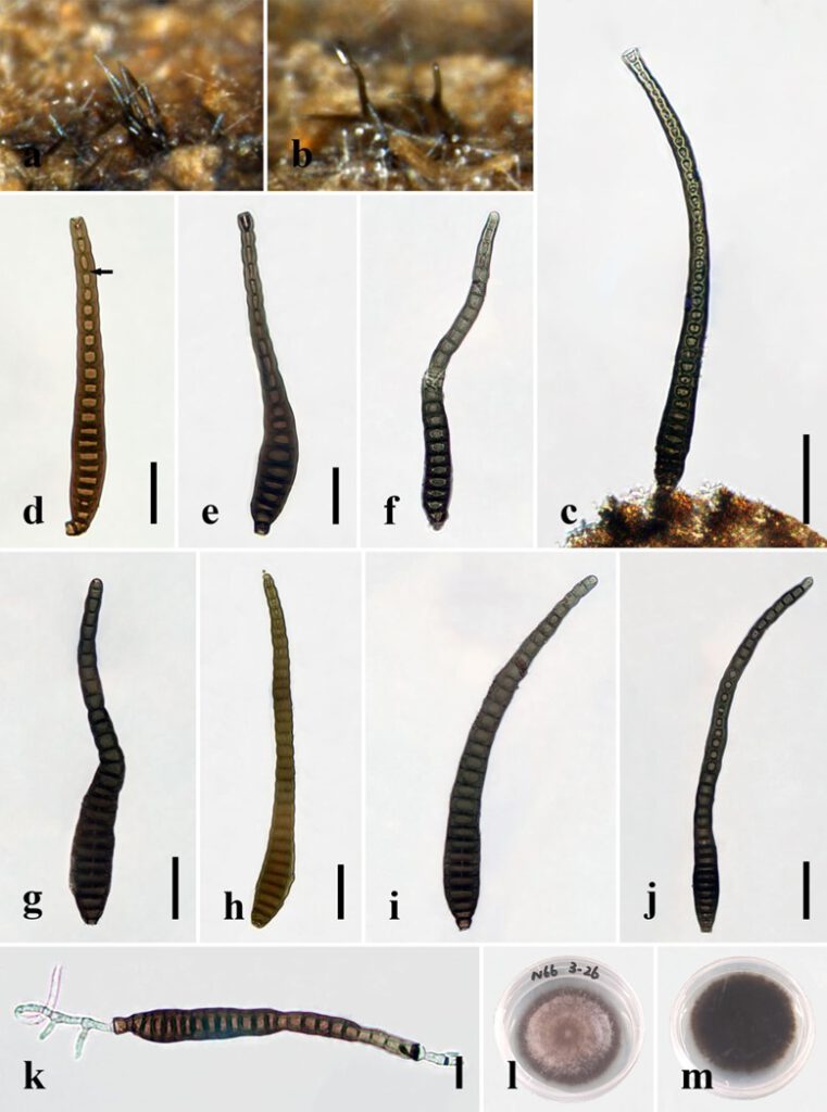
Distoseptispora crassispora R. Zhu & H. Zhang, sp. nov.
MycoBank number: MB; Index Fungorum number: IF; Facesoffungi number: FoF 12577;
Description
Saprobic on decaying wood submerged in freshwater. Sexual morph: undetermined. Asexual morph: hyphomycetous. Colonies on the substratum superficial, effuse, scattered, hairy, dark-brown. Mycelium mostly immersed, composed of branched, septate, smooth, dark-brown hyphae. Conidiophores 14–27 μm long, 6–10 μm wide, macronematous, mononematous, solitary, unbranched, septate, cylindrical, straight or flexuous, smooth, brown to dark-brown, robust at the base. Conidiogenous cells 2.5–7 μm long (x̄=4.5 μm, n=10), 5.5–8 μm wide (x̄=6 μm, n=10), monoblastic, integrated, determinate, terminal, cylindrical, dark-brown, smooth. Conidia 95–197(–214) µm long (rostrum included) (x̄=141 μm, n=30), 13–24 μm at the widest part (x̄=17 μm, n=30), 6–12 μm wide at the apex (x̄=9 μm, n=30), acrogenous, dry, obclavate, rostrate, mostly curved, 15–36(–41)-distoseptate, truncate at the base, rounded at the apex, smooth, thick-walled, brown with a green tinge.
Material examined: China, Yunnan Province, Xishuangbanna, Man Feilong Reservoir, found on dead, submerged, decaying wood of unidentified plants, 7 November 2020, Rong Zhu, N66 (HKAS 122181, holotype), ex-type living culture KUNCC 12–10726.
Distribution: China
Sequence data: ITS: OK310698 (ITS5/ITS4); LSU: OK341196 (LROR/LR5); TEF1a: OP413479 (983/2218R); RPB2: OP413473 (fRPB2-5F/fRPB2-7cR)
Notes: Multi-gene phylogenetic analyses showed that D. crassispora is a distinct species in Distoseptispora and clusters with D. chinense and D. tectonigena. Distoseptispora crassispora is morphologically similar to the latter two species in having straight to slightly curved, septate conidiophores and obclavate, dis-toseptate, rostrate, straight or slightly curved conidia. However, D. crassispora possesses shorter conidiophores (up to 27 μm vs. up to 110 μm) and wider conidia (13–24 μm vs. 10–13 μm) than D. tectonigena. Additionally, the conidia of D. tectonigena are sometimes percurrently proliferating 5–10 times at the apex, which was not observed in D. crassispora. Distoseptispora crassispora can be distinguished from D. chinense on the basis of molecular data. Comparisons of nucleotides between D. crassispora and D. chinense revealed 12 (2.3%, including four gaps) and 23 (2.6%) nucleotide differences in the ITS region and TEF1-α, respectively, which follows the generally accepted norm, according to which nucleotide differences of more than 1.5% in the ITS region are likely to indicate a new species. We therefore introduce D. crassispora as a new species in Distoseptispora.

Fig. x. Distoseptispora crassispora (holotype). (a,b) Colonies on natural substrate. (c) Conidiophores with conidium. (d–j) Conidia. The arrow in d points to a gap in the middle of the septum, which indicates the distosepta. (k) Germinated conidium. (l) Colony on PDA (from front). (m) Colony on PDA (from reverse). Scale bars: (c) 40 μm; (d–f) 20 μm; (g–j) 30 μm; (k) 2 μm.
