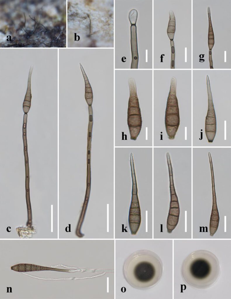
Distoseptispora aqualignicola C.X. Li & H. Zhang, sp. nov.
MycoBank number: MB; Index Fungorum number: IF; Facesoffungi number: FoF 12575;
Description
Saprobic on decaying wood submerged in freshwater. Sexual morph: undetermined. Asexual morph: hyphomycetous. Colonies on the substratum superficial, effuse, scattered, hairy, brown to dark-brown. Mycelium partly superficial, partly immersed, composed of branched, septate, smooth, hyaline to pale-brown hyphae. Conidiophores 90–190(–240) μm long (x̄=162 µm, n=15) and 5–8 μm wide (x̄=6 µm, n=15), macronematous, mononematous, unbranched, multi-septate, single or in groups of two or three, cylindrical, straight or slightly flexuous, smooth, brown, rounded at the apex. Conidiogenous cells 13–21 μm long (x̄=18 µm, n=10), 4–5.5 μm wide (x̄=4.5 µm, n=10), monoblastic, integrated, determinate, terminal, cylindrical, brown, smooth. Conidia 41–94(–104) μm long (x̄=73 µm, n=20) (rostrum included), 10.5–12.5 µm at the widest part (x̄=11.5 µm, n=20), 2–5 µm wide at the apex (x̄=3.5 µm, n=20), acrogenous, dry, obclavate, rostrate, straight or curved, 4–8-euseptate, mostly 6–7-euseptate, tapering towards the rounded apex, brown at the base, smooth, thin-walled, subhyaline to pale-brown at the apex.
Material examined: China, Sichuan Province, Yibin City, Southern Sichuan Bamboo Sea, Qicai Lake, found on dead, submerged, decaying wood of unidentified plants, 16 June 2019, Chunxue Li, S1–10 (HKAS 122184, holotype), ex-type living culture KUNCC 21–10729.
Distribution: China
Sequence data: ITS: OK341186 (ITS5/ITS4); LSU: ON400845 (LROR/LR5); TEF1a: OP413480 (983/2218R); RPB2: OP413474 (fRPB2-5F/fRPB2-7cR)
Notes: Distoseptispora aqualignicola, D. aquamyces, D. lancangjiangensis, D. meilingensis, D. suoluoensis, D. yongxiuensis and D. verrucose form a strongly supported clade (99% MLBS/1.00 BIPP) in Clade 2 of Distoseptispora (Figure 1). Morphologically, D. aqualignicola possesses smooth-walled conidia, which are distinct from the verrucose conidia of D. aquamyces, D. suoluoensis and D. verrucosa. Distoseptispora aqualignicola differs from D. meilingensis in having euseptate, thin-walled conidia as compared with the distoseptate, thick-walled conidia of D. meilingensis. Distoseptispora lancangjiangensis and D. yongxiuensis resemble D. aqualignicola with respect to their smooth-walled conidia but they are phylogenetically distinct.

Fig. x. Distoseptispora aqualignicola (holotype). (a,b) Colonies on natural substrate. (c,d) Conidiophores with conidia. (e–g) Conidiogenous cells bearing conidia. (h−m) Conidia. (n) Germinated conidium. (o) Colony on PDA (from front). (p) Colony on PDA (from reverse). Scale bars: (c,d) 30 μm; (e,h–i) 10 μm; (f,g,j–n) 20 μm.
