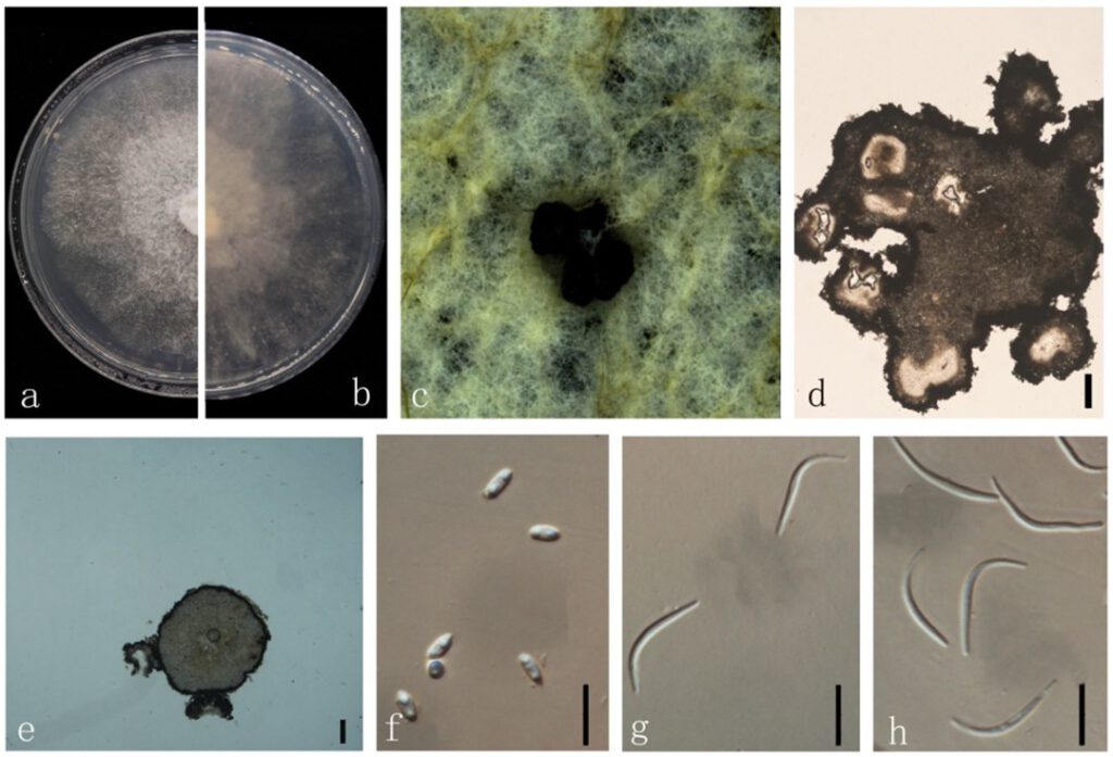
Diaporthe unshiuensis F. Huang, K.D. Hyde & Hong Y. Li, in Huang, Udayanga, Mei, et al., Fungal Biology 119(5): 344 (2015) (Figure 13)
MycoBank number: MB 810845; Index Fungorum number: IF 810845; Facesoffungi number: FoF 09422;
Endophytic on Morinda officinalis stem. Sexual morph: not observed. Asexual morph: Pycnidia 350×300 μm, globose, subglobose or irregular, bark brown, parenchymatous walls. Conidial masses produced as black droplets extruding through the ostioles. Pycnidia wall consisting of sevea layers of medium transparent textura globosa-angularis. Pycnidia wall consisting of sevea layers of medium transparent textura globosa-angularis, conidial cirrus extruding from ostiole appearing as black droplets. Conidiophore cylindrical, wide at the base, hyaline, simple. Alpha conidia 5–10 × 2–3 μm (`x = 6 ± 0.8 × 3 ± 0.3 μm), ellipsoidal or clavate, base truncate, aseptate, smooth, biguttulate. Beta conidia 20–30 × 1–3 μm (`x = 25 ± 3 × 2 ± 0.4 μm), hyaline, linear, curved, hook-shaped.
Culture characteristics: Colonies on PDA reach 85 mm diam. after 9 days. Surface white and turning to grey with ageing, reverse white to grey with dark grey at the centre.
Material examined: China, Guangdong Province, Zhaoqing, isolated from healthy root of Morinda officinalis. June 2020, W. Guo, dried culture (ZHKU 22-0037), and living culture (ZHKUCC 22-0051–53).
Habitat and host: Carya illinoinensis, Citrus sp., Citrus unshiu, Fortunella margarita, Glycine max and Vitis vinifera [45].
Known distribution: China, Louisiana [45].

Figure 13. Diaporthe unshiuensis (ZHKUCC 22-0051) (a) upper view of colonies on PDA; (b) Reverse view of colonies on PDA; (c) Conidiomata sporulating on PDA; (d, e) Section of pycnidia; (f) Alpha conidia; (g, h) Beta conidia. Scale bars: (d,e) =100 µm; (f–h) =10 µm.
