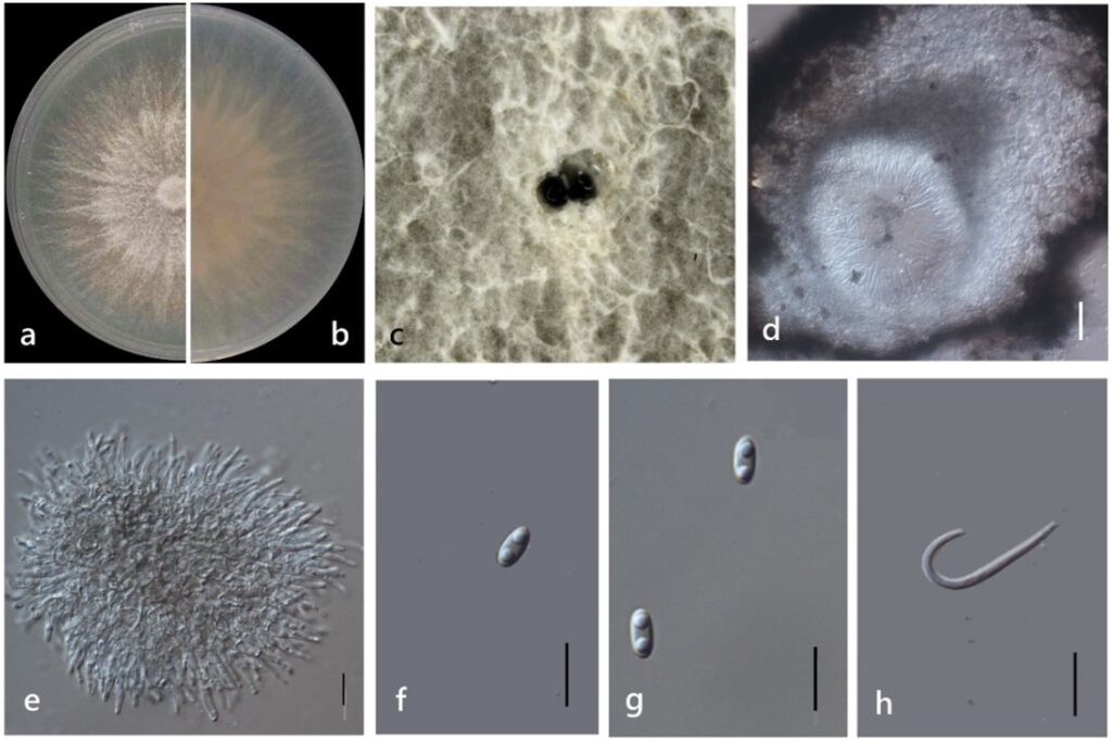
Diaporthe morindendophytica M. Luo, W. Guo, Manawas., M. P. Zhao, K. D. Hyde, & C. P. You, sp. nov. (Figure 11)
MycoBank number: MB; Index Fungorum number: IF; Facesoffungi number: FoF 11355;
Etymology: In reference to its endophytic nature in the host of Morinda officinalis.
Holotype: ZHKUCC 22-0069
Endophytic on Morinda officinalis. Sexual morph: not observed. Asexual morph: Pycnidia 40–200 × 30–130 μm (`x = 120 ± 450 μm× 70 ± 20 μm), oblate, subglobose or irregularly shaped, single or multiple cavities. Pycnidial wall consisting of sevearal layers of medium transparent textura globosa-angularis. Conidial masses produced as black droplets extruding through the ostioles.Conidiogenus cells hyaline, phialidic. Alpha conidia 5–10 × 2–3 μm (`x = 6 ± 0.5 μm× 3 ± 0.3 μm), hyaline, ellipsoid, torque circular or lanceolate, blunt at both ends or slightly pointed at one end, biguttulate. Beta conidia 20–30 × 1–2 μm (`x = 20 ± 3 μm× 2 ± 0.2 μm), hyaline, linear, curved.
Culture characteristics: Colonies on PDA reach 85 mm diam. after 4 days. White cotton flocculent aerial hyphae, turned grayish-black or yellow. Reverse yellowish-brown, with a large number of black dots.
Material examined: China, Guangdong Province, Zhaoqing, isolated from a healthy stem of Morinda officinalis. June 2020, W. Guo, dried culture (ZHKU 22-0042), and living culture (ZHKUCC 22-0069 ex-type and ZHKUCC 22-0070, 22-0071 ex-paratype).
Habitat and host: Healthy stem of Morinda officinalis.
Known distribution: China (Zhaoqing, Guangdong Province).

Figure 11. Diaporthe morindendophytica (ZHKUCC 22-0069) (a) Upper view of colonies on PDA; (b) Reverse view of colonies on PDA; (c) Pycnidia sporulating on PDA; (d) Section of pycnidia; (e) Conidiogenous cells; (f,g) Alpha conidia; (h) Beta conidia. Scale bars: (d) =100 µm; (e–h) =10 µm.
