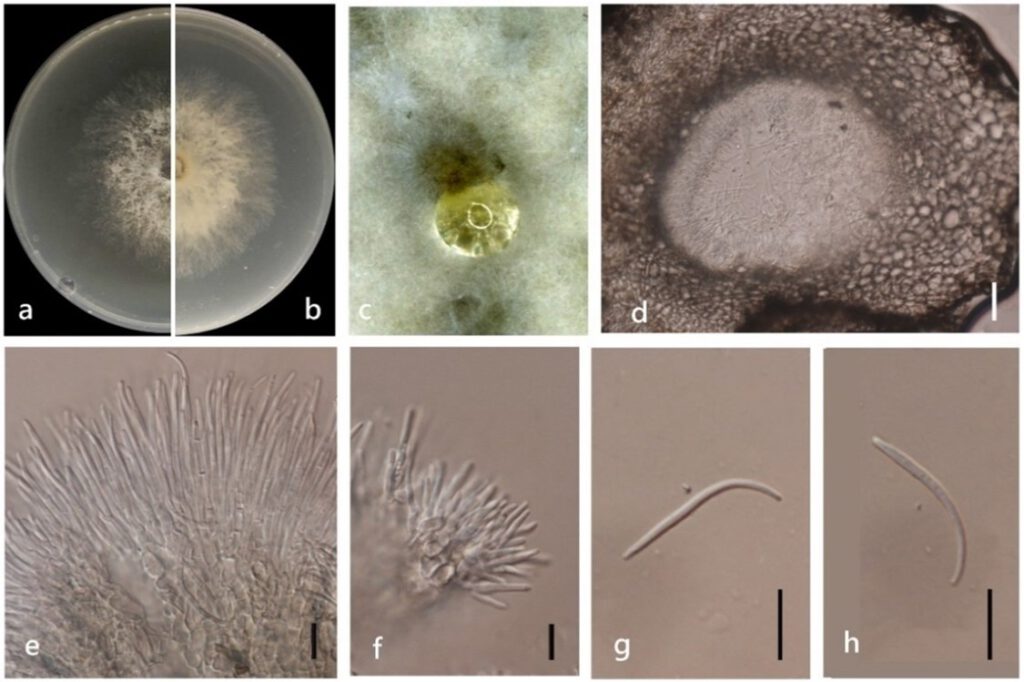
Diaporthe xishuangbanica Y.H. Gao & L. Cai, in Gao, Liu, Duan, et al., IMA Fungus 8(1): 179 (2017) (Figure 14)
MycoBank number: MB 820685; Index Fungorum number: IF 820685; Facesoffungi number: FoF 11356;
Endophytic on Morinda officinalis root and stem. Sexual morph: not observed. Asexual morph: Pycnidia 30 – 360 × 20 – 230 μm (`x = 119 ± 90 μm× 72 ± 60 μm), oblate, subglobose or irregularly shaped, single or multiple cavities. Conidial masses produced as transparent droplets extruding through the ostioles. Pycnidia wall consisting of sevea layers of medium transparent textura globosa-angularis. Conidial masses produced as transparent droplets extruding through the ostioles. Conidiophores hyaline, branched, septate. Conidiogenus cells grow from the conidia, hyaline, phialidic. Beta conidia 20 – 30 × 1 – 2 μm (`x = 27 ± 3 μm× 2 ± 0.3 μm), hyaline, linear, curved. Alpha conidia and gamma conidia not observed.
Culture characteristics: Colonies on PDA reach 85 mm diam. after 5 days. White cotton flocculent aerial mycelium in the center, surrounding sparse hyphae. At 15 days, white cotton flocculent aerial hyphae and a few black dots appear, reddish pigments are produced on the back side of the colony. At 30 days, the black dots increased and the fruiting body was produced.
Material examined: China, Guangdong Province, Zhaoqing, isolated from healthy root of Morinda officinalis. June 2020, W. Guo, dried culture (ZHKU 22-0038), and living culture (ZHKUCC 22-0054, 22-0055).
Habitat and host: Camellia sinensis [8].
Known distribution: China [8].

Figure 14. Diaporthe xishuangbanica (ZHKUCC 22-0054) (a) upper view of colonies on PDA; (b) Reverse view of colonies on PDA; (c) Pycnidia with conidial droplets on PDA; (d) Section of pycnidia; (e,f) Conidiogenous cells; (g,h) Beta conidia. Scale bars: (d) =100 µm; (e–h) =10 µm.
