Botryosphaeria minutispermatia Ariyawansa, K.D. Hyde, Z.Y. Liu, sp. nov., Index Fungorum number: IF 552252
Etymology:—In reference to the tiny spermatia produced in culture.
Saprobic on dead wood. Sexual morph: Ascostromata embedded in the host, becoming partially erumpent at maturity, mostly solitary. Ascomata 190−300 μm wide, forming a botryose aggregate, globose with a central ostiole, papillate, brown to black. Peridium 50–100 μm (x̅=72 μm, n=10) wide, consisting of 5–15 regions with 2–4 layers of hyaline cells lining the locule. Hamathecium of dense, 2–4 μm wide, filiform, septate, rarely branched pseudoparaphyses, constricted at the septa. Asci 70–90×15–18 μm (x̅=81×17 μm, n=20), 8–spored, bitunicate, fissitunicate, broadly clavate, with short, broad pedicel with well-developed ocular chamber, forming between pseudoparaphyses. Ascospores 28–31×8–10μm (x̅=30×9 μm, n=40), biseriate, fusoid to ovoid, hyaline, thick-walled, aseptate with tapered ends. Asexual morph: Conidiogenous cells 5–9×1–3 μm, holoblastic, subcylindrical, hyaline. Conidia 8–14×3–4 μm (x̅=13×3.5 μm, n=40), irregularly fusiform, hyaline, aseptate when young, becoming 1-septate at maturity, smooth-walled, with granular contents. Spermatia 1–5×0.2–1 μm, hyaline, rod-shaped.
Culture characteristics: Colonies on PDA growing rapidly, reaching 10 cm diam. within 7 days at room temperature, aerial mycelium at first white becoming dark grey to black.
Material examined:—CHINA, Guizhou Province, Guiyang City, Huaxi District, Guizhou Academy of Agricultural Sciences garden, on dead wood, 14 December 2015, HA Ariyawansa (GZAAS 16-0009!, holotype), ex-type living culture (GZCC 16-0013); ibid. (MFLUCC, isotype), living culture, MFLUCC.
Additional material examined:—CHINA, Guizhou Province, Guiyang City, Huaxi District, Guizhou Academy of Agricultural Sciences garden, on dead wood, 20 January 2016, HA Ariyawansa (GZAAS 16-0010!, paratype), living culture, GZCC 16-0014.
Notes:— Botryosphaeria minutispermatia fits in the generic concept of Botryosphaeria in having erumpent ascomata, bitunicate, clavate asci, with a well-developed ocular chamber, hyaline pseudoparaphyses and hyaline, fusoid to ellipsoid or ovoid, ascospores and fusicoccum-like asexual morph produce in culture (Liu et al. 2012, Phillips et al. 2013, Zhou et al. 2016). The hyaline conidia with a single septum at maturity, the narrow spermatia formed in the culture and the relatively smaller ascomata with large hyaline ascospores, readily distinguish B. minutispermatia from the other species in the genus. Botryosphaeria minutispermatia and the generic type of Botryosphaeria, B. dothidea are similar in having erumpent ascomata, a peridium comprising several regions of 2–4 layers of hyaline cells lining the locule, filiform pseudoparaphyses, clavate asci forming between pseudoparaphyses and fusoid to ovoid ascospores and a fusicoccum-like asexual morph (Liu et al. 2012, Phillips et al. 2013, Zhou et al. 2016). Differences are in the size of the ascomata in ascostromata (170–300 μm versus 200–500 μm in ascostromata), asci (70–90×15–18 μm vs. 63−125×16–20 μm) and ascospores (28–31×8–10μm vs. 19–24×7–8μm) separate the species (Liu et al. 2012, Phillips et al. 2013, Zhou et al. 2016). Furthermore, Botryosphaeria minutispermatia differs from B. dothidea in having smaller conidia (8–14×3–4 μm versus 18–20×4–5 μm) and smaller spermatia (1–4×0.2–1 μm versus 3–6×1.5–2 μm). Based on both morphology and phylogeny, Botryosphaeria minutispermatia shares similarities with B. sinensia (Zhou et al. 2016), but they differ in the size of the ascomata in ascostromata and the sizes of the asci, ascospores, conidia and spermatia.
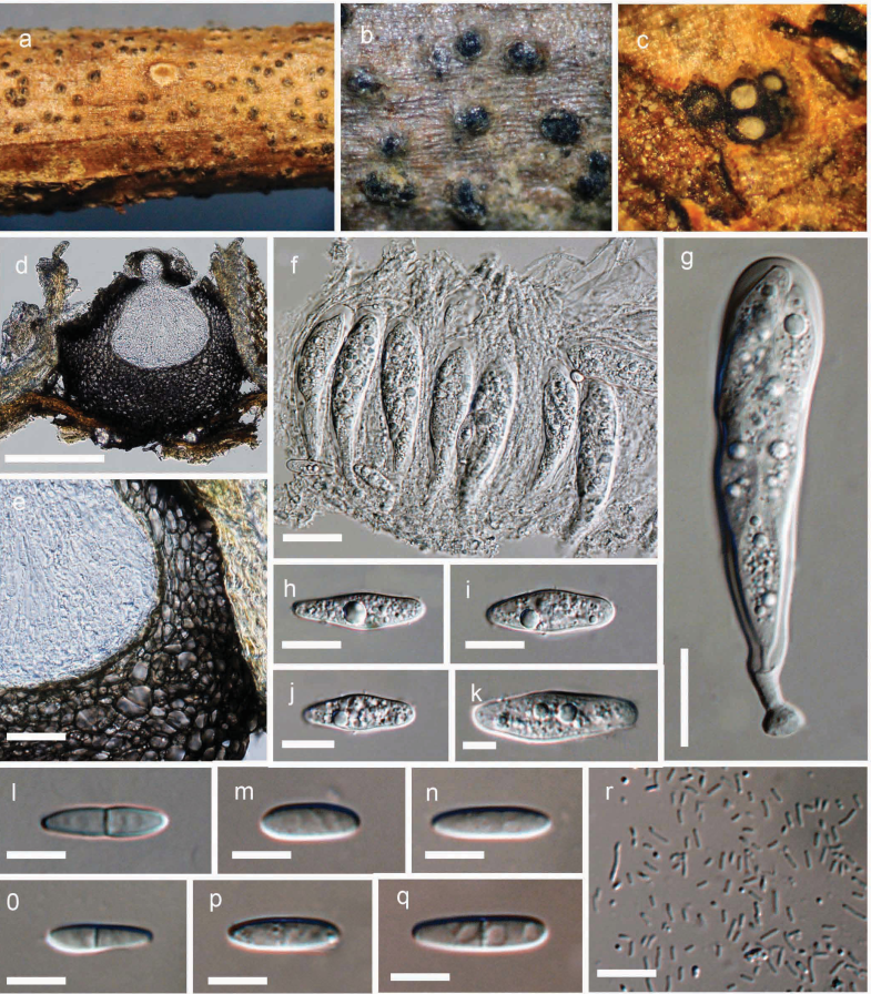
FIGURE. Botryosphaeria minutispermatia (holotype). a Ascostromata on host substrate. b Close up of the ascostromata c Botryose arrangement of ascomata in ascostromata. The tops of two have been removed to show the typical white centrum content. d Section through a mature ascomata. e Close up of the peridium. f Hamathecium of dense filiform, septate pseudoparaphyses with asci. g Mature ascus with hyaline spores. h–k Fusoid to ovoid, hyaline, aseptate ascospores. l–q Mature and immature conidia produced in culture. r Spermatia. Scale bars: d = 150 μm, e = 20 μm, f = 15 μm, g, l-q = 5 μm, h-k = 10 μm, r = 10 μ.