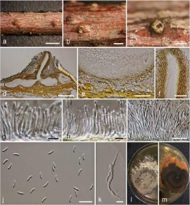
Cytospora ulmi Norphanphoun, Bulgakov, T.C. Wen & K.D. Hyde, sp. nov.,Index Fungorum number: IF552614
Etymology: Name refers to the host genus Ulmus
Holotype: MFLU 15-1910
Associatd with twigs and branches of Ulmus minor. Sexual morph: Undetermined. Asexual morph: Conidiomata 980–1200 × 500–600 μm diameter, semi-immersed in host tissue, solitary, erumpent, scattered, discoid, circular to ovoid, with 4–6 locules, with long ostiolar neck. Ostioles 410–450 μm, at the same level as the disc surface. Peridium comprising a few to several layers of cells of textura angularis, with inner most layer thin, pale brown. Conidiophores branched, reduced to conidiogenous cells. Conidiogenous cells blastic, enteroblastic, phialidic, hyaline, smooth-walled. Conidia (4.8–)5.4–6.4 × 1.3–1.5(–1.6) μm (x̅ = 5.4 × 1.4 μm, n = 30), unicellular, elongate-allantoid, hyaline, smooth-walled.
Culture characteristics: Colonies on MEA, reaching 6 cm diameter after 7 days at 25 °C, producing dense mycelium, circular, margin rough, white, lacking aerial mycelium.
Material examined: RUSSIA, Rostov Region, Krasnosulinsky District, Donskoye forestry, ravine forest, on dead and dying branches (necrotrophic/saprobic) of Ulmus minor Mill., 28 June 2015, T. Bulgakov, T-521 (MFLU 15-2225, holotype, KUN, isotype), ex-type living culture, MFLUCC 15-0863, KUMCC.
Notes: Phylogenetic analysis of four combined gene loci indicate that Cytospora ulmi (MFLUCC 15-0863), forms a distinct branch as a sister taxon to C. ribis Ehrenb. (284-2) isolated from Thuja orientalis L., C. rosarum Grev. (218) isolated from Rosa canina L. and C. tanaitica Norphanphoun, Bulgakov & Hyde (MFLUCC 14-1057) isolated from Betula pubescens Ehrh. (Fig. 2) (Adams et al. 2002, Ariyawansa et al. 2015, Fotouhifar et al. 2010).
Cytospora ulmi is most similar to C. rosarum in having long conidia ((4.8–)5.4–6.4 μm versus 4.5–6.7 μm), but has narrower conidia (1.3–1.5(–1.6) μm versus 0.9–1.1 μm). Cytospora ribis and C. tanaitica are similar to C. ulmi in having multi-loculate conidiomata. However, C. ulmi (5.4 × 1.4 μm) has larger conidia than C. ribis (4 × 1 μm) and C. tanaitica (3.4 × 0.7 μm) (Adams et al. 2002, Fotouhifar et al. 2010, Ariyawansa et al. 2015).
FIG: Cytospora ulmi (MFLU 15-2225, holotype). a Stromatal habit in wood. b Fruiting bodies on substrate. c Surface of fruiting bodies. d Cross section of the stroma showing conidiomata. e Peridium. f Ostiolar neck. g–i Conidiogenous cells with attached conidia. j Mature conidia. k Germinating spore. l, m Colonies on MEA (l-from above, m-from below). Scale bars: a = 2000 μm, b= 500 μm, c, d = 200 μm, e, f = 50 μm, g, h = 5 μm, i, j = 10 μm, k = 20 μm.

