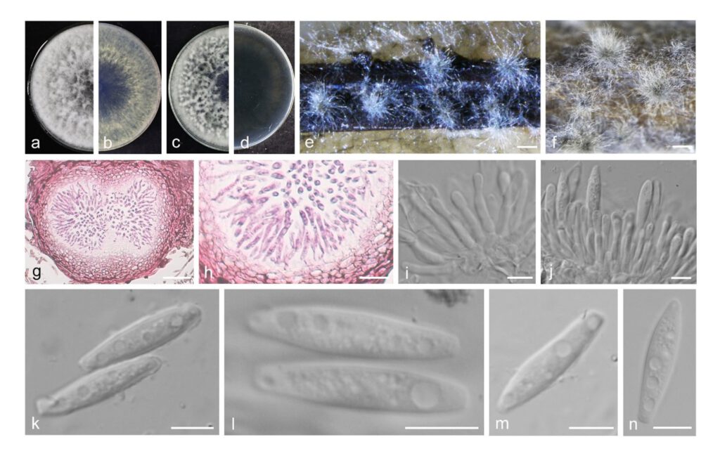
Botryosphaeria eriobotryae Yanfen Wang & M. Zhang, sp. nov. Fig. 8
MycoBank number: MB 843700; Index Fungorum number: IF 843700; Facesoffungi number: FoF 12930;
Etymology: Name reflects the host Eriobotrya japonica.
Holotype: HMAS351942
Description
The sexual stage was not observed. Conidiomata pycnidial, produced on pine needles on WA with 2–3 wk, solitary, globose to ovoid, dark brown to black, embedded in needle tissue, semi-immersed to superficial, unilocular, with a central ostiole, up to 383 μm high, up to 542 μm wide. Conidiophores absent. Conidiogenous cells holoblastic, discrete, hyaline, cylindrical to lageniform, phialidic with periclinal thickening, (8.5–)10–15(–17) × (2–)3–3(–2) μm. Paraphyses not seen. Conidia hyaline, thin-walled, smooth with granular contents, aseptate, narrowly fusiform, base subtruncate to bluntly rounded, (19.5–)21–23(–24.5) × 4–5 μm (av. = 22.3 × 4.6 μm, n = 50; L/W = 4.8).
Material examined: CHINA, Henan Province, Zhengzhou City, from stems of loquat, fruiting structures induced on needles of Pinus sp. on water agar, 15 May 2021, Y.F. Wang & M. Zhang (holotype HMAS 351942, culture ex-type CDZM 216 = CGMCC3.20835); ibid., culture CDZM 211, CDZM 217, CDZM 219 and CDZM 369.
Sequence data: ITS: OL839285 (ITS1/ITS4); EF1a: OM228523 (728F/986R); TUB2: OM228568 (Bt2A/Bt2B)
Notes: Botryosphaeria eriobotryae forms an independent clade in the Botryosphaeria, with five isolates clustering separately in a well-supported clade (BI/ML = 1/100). Conidia of B. eriobotryae differs from B. qingyuanensis in having longer and narrower conidia (22.3 × 4.6 vs 22 × 6.2 μm) (Li et al. 2018). Moreover, B. eriobotryae is most closely related to B. qingyuanensis, but easily distinguished from this species in ITS, tef1 and tub2 loci by 13 nucleotide differences in concatenated alignment (one in ITS, four in tef1 and eight in tub2).

Fig. 8 Botryosphaeria eriobotryae. a–d. colony growing on front and back of PDA (a, b) and MEA (c, d); e–f. conidiomata on PNA; g–h. section view of conidiomata; i–j. conidiogenous cells and developing conidia; k–n. conidia. — Scale bars: e–f = 500 μm; g = 50 μm; h = 20 μm; i–n = 10 μm.
