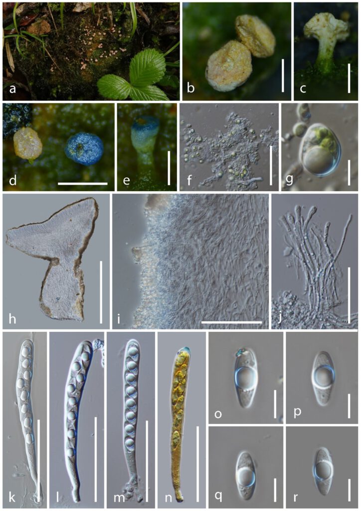
Dibaeis jingdongensis C.J.Y. Li & K.D. Hyde sp. nov
MycoBank number: MB; Index Fungorum number: IF; Facesoffungi number: FoF 12928;
Thallus crustose, green, unevenly lumpy, mostly wrinkled when fresh and dry, photobiont a unicellular green alga, ellipsoid cells with one narrow rounded end, 10–15 × 8–14 μm (x̅ = 13 × 11 mm, n = 20). Apothecia cup-shaped when fresh and dry, 1–1.5 mm wide × 0.7–1.5 mm high (x̅ = 1.3 × 1.0 mm, n = 20) when dry, scattered, superficial. Disc flat and circular, pink when fresh, pink to whitish pink, wrinkled with yellow refraction particles on surface when dry. Receptacle 0.3–0.7 mm high, smooth and flat when fresh, wrinkled and rough when dry, concolorous to the disc. Stipe 0.1–0.3 mm wide × 0.8–1.0 mm high (x̅ = 0.2 × 0.9 mm, n = 20), smooth, whitish pink, covered with unicellular green alga. Excipulum 35–55 μm, thin, comprised of irregular yellowish brown refraction particles. Hypothecium 160–260 μm, thick, comprising densely hyaline cells of textura intricata, hyphae 3–5 μm, thin-walled, septate, branched and non-gelatinous. Hymenium 140–160 μm, hyaline, covering the same materials to the Ectal excipulum on the outer layer, turning greenish blue in Melzer’s reagent. Paraphyses 3.5–4.7 μm (x̅ = 4.2 μm, n = 25) wide at tips, numerous, filiform, hyaline, obtuse and swollen at the apex, branched, aseptate, rough, guttulate, exceeding the asci in length. Asci 92–127 × 9–13 μm (x̅ = 108 × 11 μm, n = 50), 8-spored, Icmadophila-type, narrowly cylindric, apex rounded, slightly thicken with amyloid on the outmost part, tapering to subtruncate base. Ascospores (15.2–)15.8–20.1(–22.2) × (5.6–)5.8–7.3(–7.5) μm (x̅ = 18.0 × 6.4 μm, n = 100), Q = 2.2–3.4(–3.8) μm, Qm = 2.8 ± 0.1 μm, uniseriate, sometimes overlapping, ellipsoidal, tapering to narrowly rounded at one end, hyaline, thick-walled, smooth, aseptate with a big guttule. Pycnidia not found.

Figure 1. Dibaeis jingdongensis (HKAS 125549, holotype). a Fresh ascomata on natural habitat. b Dried ascomata on substrate. d–e Rehydrated ascomata and pigmented in Meltzer’s reagent. f–g Cells of photobiont. h Vertical section of ascomata. i Excipulum. j Paraphyses. k-n Asci (Asci in Meltzer’s reagent). o–p Ascospores. Scale bar: b, h = 1000 μm, c = 1200 μm, d = 2000 μm, e = 800 μm, f = 70 μm, g = 6 μm, i = 100 μm, j = 40 μm, k–n = 60 μm, o–r = 8 μm.
