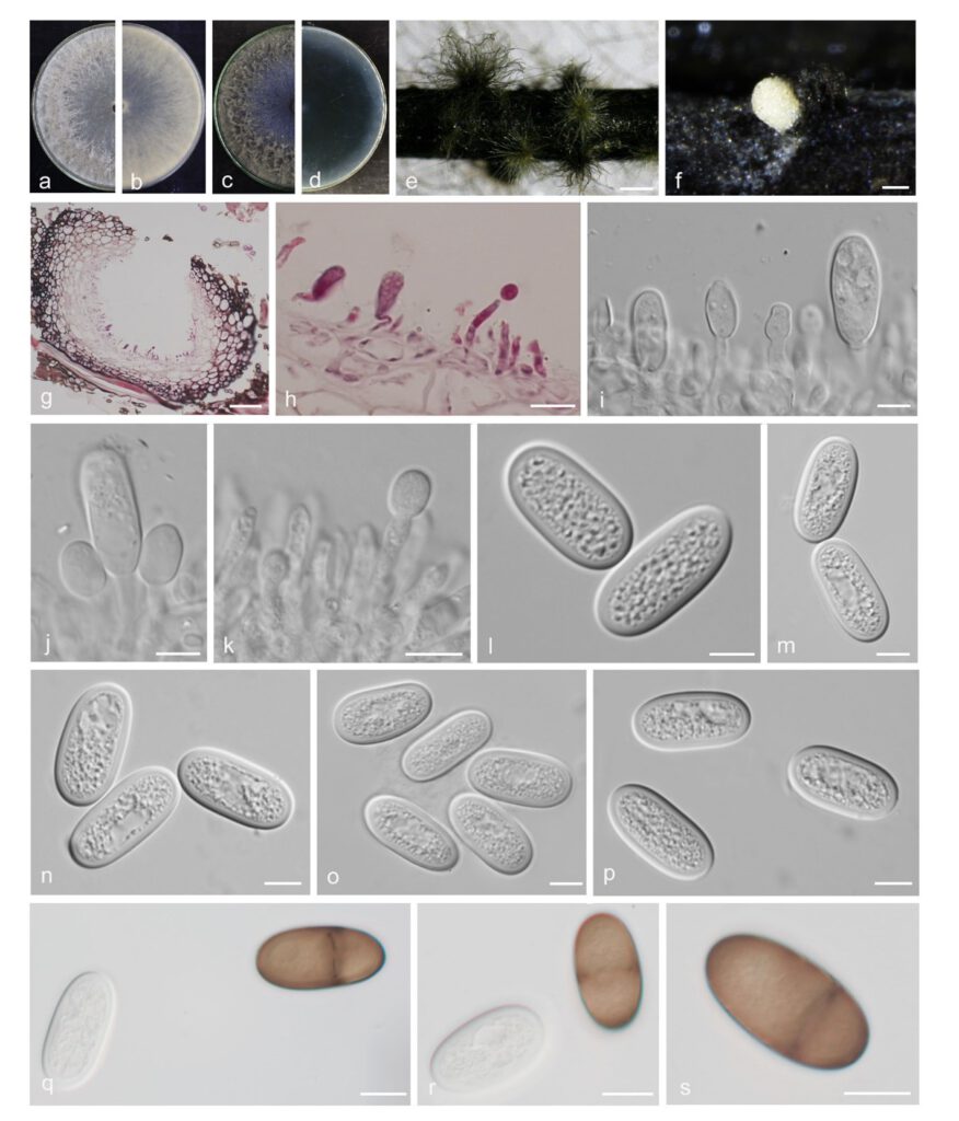
Diplodia pruni-persicae Yanfen Wang & M. Zhang, sp. nov. Fig. 11
MycoBank number: MB 843628; Index Fungorum number: IF 843628; Facesoffungi number: FoF 12931;
Etymology: Name refers to Prunus persica, the host from which this fungus was collected.
Holotype: HMAS 351913
Description
The sexual stage was not observed. Conidiomata pycnidial, produced on pine needles on WA within 2–3 wk, solitary or aggregated, individual conidiomata globose, superficial, covered with hyphal hairs, uniloculate, thick-walled, exuding conidia in a yellow mucoid mass, up to 385 μm high, up to 473 μm wide. Conidiogenous cells hyaline, thin-walled, smooth, cylindrical, discrete, holoblastic, proliferating percurrently to form two or three distinct annellations or proliferating internally giving rise to periclinal thickenings, (8–)9–11.5 × (2.5–)3–3.5 μm. Paraphyses absent. Conidia oblong, both ends broadly rounded, initially hyaline, aseptate, becoming dark brown and 1-septate with age, thick-walled, smooth, (22–)23.5–25.5(–26.5) × (10–)11–12.5(–13) μm (av. = 24.5 × 12 μm, n = 50; L/W = 2.0).
Material examined: CHINA, Henan Province, Xinyang City, from branches of peach, fruiting structures induced on needles of Pinus sp. on water agar, 1 Jul. 2021, Y.F. Wang & M. Zhang (holotype HMAS 351913, culture ex-type CDZM 921 = CGMCC 3.20837; CDZM 922); Xinyang City, from branches of loquat, 1 Jul. 2021, Y.F. Wang & M. Zhang (culture CDZM 432 and CDZM 434).
Sequence data: ITS: OL863126 (ITS1/ITS4); EF1a: OM228566 (728F/986R); TUB2: OM228612 (Bt2A/Bt2B)
Notes: Diplodia pruni-persicae forms a distinct clade, and differed with the closely related species (D. neojuniperi and D. mutila) on morphology. Conidia of D. pruni-persicae are longer and wider than D. neojuniperi (24.5 × 12 vs 23.9 × 14.8 µm) (Trakunyingcharoen et al. 2015), but shorter and narrower than D. mutila (24.5 × 12 vs 25.4 × 13.4) (Slippers et al. 2014). Moreover, conidia of D. pruni-persicae become dark brown with age and longitudinal striations, while those of D. mutila are rare. In terms of the nucleotide comparison, D. pruni-persicae differed from these species by nucleotide differences in ITS (D. neojuniperi: 3 bp and 3 indel, D. mutila: 2 bp and 3 indel), tef1 (D. neojuniperi: 3 bp and 15 indel, D. mutila: 1 bp) and tub2 loci (tub2 is not available to D. neojuniperi, D. mutila: 1 bp and 3 indel).

Fig. 11 Diplodia pruni-persicae. a–d. colony growing on front and back of PDA (a, b) and MEA (c, d); e–f. conidiomata on PNA; g–h. section view of conidiomata; i–k. conidiogenous cells; l–p. aseptate, hyaline conidia; q–r. hyaline, aseptate and brown, 1-septate mature conidia; s. brown, 1-septate mature conidia. — Scale bars: e = 500 μm; f = 200 μm; g = 50 μm; h–s = 10 μm.
