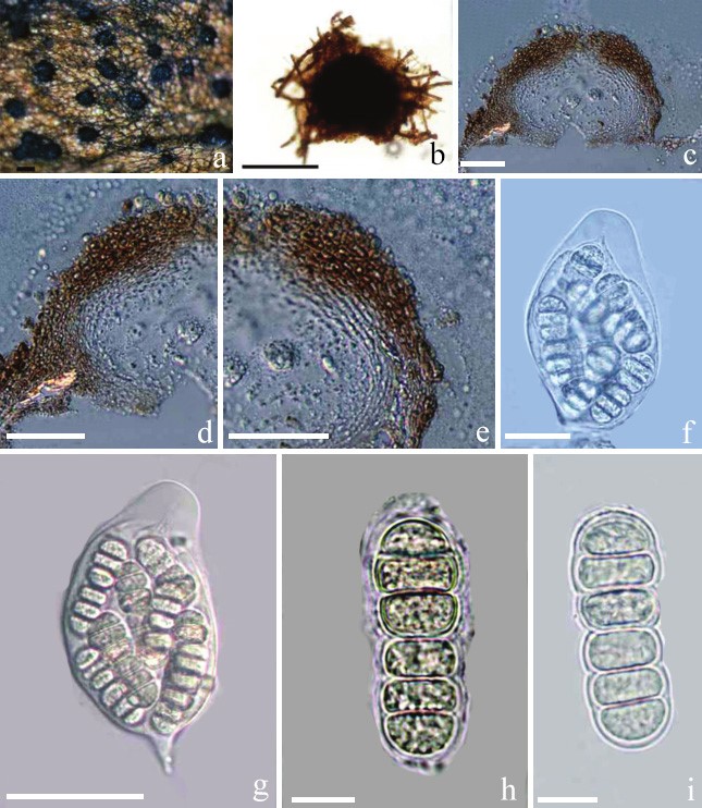
Beelia suttoniae F. Stevens & R.W. Ryan, Bulletin of the Bernice P. Bishop Museum, Honolulu, Hawaii 19: 71 (1925), Figure 5
MycoBank number: MB 200511; Index Fungorum number: IF 200511; Facesoffungi number: FoF 10331;
Saprobic on the surface of leaves of Suttonia lanaiensis. Sexual morph: Ascomata 190–210 µm wide, 115–133 µm high, scattered, superficial, immersed in the darkened mycelial substrate, globose to subglobose, black, easily removed, ostiolate. Ostiole open, periphysate. Wall of ascoma 25–30 µm wide, up to 39 µm wide at the apex, 20 µm wide at the base, comprising multi-layers, externally comprising pigmented, dark brown, thick-walled cells of textura globulosa, with inner layer thinner, composed of lightly pigmented to hyaline, thin-walled cells of textura angularis. Hamathecium lacking paraphyses. Asci 70–89 × 45–55 µm (x̅ = 84.3 × 51.2 µm, n = 20), 8-spored, bitunicate, broadly ellipsoidal, obovate to saccate, thick-walled, with a small pointed pedicle, with an ocular chamber. Ascospores 38–45 × 13–18 µm (x̅ = 42.8 × 14.6 µm, n = 20), irregularly arranged, cylindrical, hyaline, 5-septate, slightly constricted at each septum, central septum strongly constricted and upper part wider, smooth-walled, with a narrow mucilage sheath. Asexual morph: Undetermined.
Material examined: USA, Hawaii, on leaves of Suttonia lanaiensis Mez (Myrsinaceae), 1925, Lanai, no. 421, leg. Munro (BISH 499845, syntype).

Figure 5 Beelia suttoniae (BISH 499845, syntype). a Appearance of ascomata on host leaf with darkened mycelium. b Squash of ascoma in water. c–e Vertical section through ascoma. f, g Asci with ascospores. h, i Ascospores. Scale bars: b = 100 µm, c–e, g = 50 µm, f = 25 µm, h= i = 10 µm.
