Aspergillus quadrilineatus Thom & Raper, Mycologia 31: 660. (1939). Figs 2 & 3.
MycoBank number: MB 275888; Index Fungorum number: IF 275888; Facesoffungi number: FoF 10610;
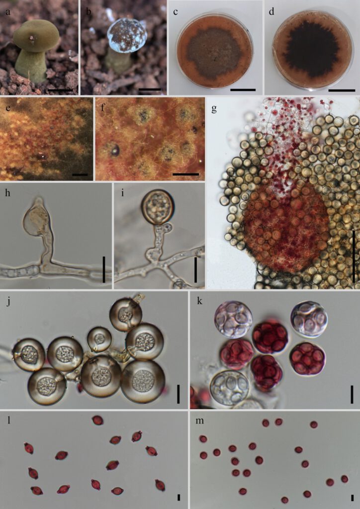
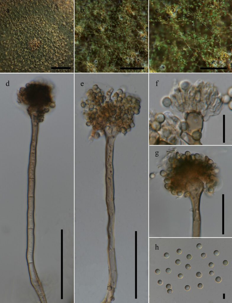
Fungicolous on the fruiting body of Phlebopus spongiosus Pham and Har. Takah. Sexual morph on PDA: Ascomata 190–310 × 160–300 μm (x̅ = 227.6 × 221.6 μm, n = 5), cleistothecial, developing throughout the colony, reddish-brown, globose to subglobose, surrounded by numerous Hülle cells; Hülle cells 12.5–23 × 11.5–22 μm (x̅ = 17.5 × 17.3 μm, n = 30), hyaline to brown, globose to ovoid. Asci 9–11 × 8–11 μm (x̅ = 10.1 × 9.9 μm, n = 15), 8-spored, globose to subglobose. Ascospores 3–4 × 3–4 μm (x̅ = 3.8 × 3.4 μm, n = 40), orange to reddish-brown, globose to subglobose in surface view, spore bodies smooth, lenticular in lateral view, with two plaited equatorial crests about 0.5-1 μm in width paralleled by a secondary narrower pair which are sometimes indistinct, crests are entire, defective or with irregular protuberance. Asexual morph on host: Mycelium powdery mass, white to green; sporulation light green to dark green. PDA: Conidiophores 80–165 × 3–6 μm (x̅ = 122.2 × 4.3 μm, n = 15) macronematous, mononematous, erect, simple, with septate to aseptate stipes, pale brown; Vesicles 9–12 μm (x̅ = 10.6 μm, n = 15) wide, pale brown, globose to subglobose; Metulae 3–4.5 × 1–2 μm (x̅ = 3.7 × 1.5 μm, n = 20) hyaline; Phialides 4.5–6 × 1–2 μm (x̅ = 5.7 × 1.4 μm, n = 20) hyaline, flask-shaped; Conidia 2.5–4 × 2–4 μm (x̅ = 3.2 × 3.3 μm, n = 50) slightly echinulate, globose to subglobose, hyaline to light green.
Colony characters: on PDA, 25 °C, 14 days: Colonies moderately deep, marginally sulcate; margins slightly irregular; mycelium buff and white, later turns to reddish-brown; texture floccose; sporulation sparse; soluble pigments absent; exudates clear droplets; reverse dark brown.
Material examined: THAILAND, Chiang Rai Province, growing on the fruiting body of Phlebopus spongiosus cultivated in soil, 29 November 2019, Bhavesh Raghoonundon, AJ 044 (MFLU xxxx), living cultures MFLUCC 21-0165 and MFLUCC 21-0166.
GenBank accession numbers: MFLUCC 21-0165: ITS: OL615082; tub2: OL625674. MFLUCC 21-0166: ITS: OL615089; tub2: OL625675.
Notes: Aspergillus quadrilineatus strains MFLUCC 21-0165 and MFLUCC 21-0166 reported in this study share similar sexual and asexual morphologies with those observed from the culture of the neotype of A. quadrilineatus (NRRL201) described by Chen et al (2016). The ascomata, Hülle cells, ascospores, vesicles, metulae and phialides of MFLU xxxx are comparatively smaller than those of A. quadrilineatus described in Chen et al. (2016) as shown in Table 2. These dimensional differences occurred may be due to the variations of agar medium used to obtain the culture.
During this study, all four loci namely, ITS, tub2, cam and rpb2 were amplified and sequenced using methods and primers as previously described in Houbraken & Samson (2011) and Samson et al (2014). However, only the ITS and tub2 sequence results were obtained successfully. There were no base pair differences of ITS and tub2 sequences between the neotype of A. quadrilineatus (NRRL201) described by Chen et al (2016) and Aspergillus quadrilineatus strains MFLUCC 21-0165 and MFLUCC 21-0166 reported in this study.
Table 2: The comparison of the sizes of fungal structures
| Fungal structure | Chen et al. (2016) | This study |
| Ascomata | 100–700 µm | 190–310 × 160–300 µm |
| Hülle cells | 10–24 μm | 12.5–23 × 11.5–22 μm |
| Ascospores | 4–4.5 × 3–4.5 µm | 3–4 × 3–4 μm |
| Vesicles | 10–13 µm | 9–12 μm |
| Metulae | 5–7 × 2–4.5 μm | 3–4.5 × 1–2 μm |
| Phialides | 5–7 × 2–4 µm | 4.5–6 × 1–2 μm |

Figure 1 – Sexual morph of Aspergillus quadrilineatus (MFLUCC 21-0165). a. Healthy mushroom, Phlebopus spongiosus growing in soil. b. The mushroom affected by Aspergillus quadrilineatus. c,d. Colony on PDA. e,f. Cleistothecial ascomata formation in the colony on PDA. g. Cleistothecium covered with Hülle cells. h,i. Formation of Hülle cells. j. Hülle cells. k. Immature and mature asci. l. Side view of ascospores. m. Surface view of ascospores. Scale bars: a,b = 2 cm, c,d = 30 mm, e = 300 μm, f = 500 μm, g = 100 μm, h–j = 10 μm, k = 5 μm, l, m = 3 μm.

Figure 2 – Asexual morph of Aspergillus quadrilineatus (MFLUCC 21-0165). a. Colony on PDA. b,c. Sporulation on PDA. d,e. Mononematous conidiophores with conidia. f,g. Apex of conidiogenous cell. h. Conidia. Scale bars: a = 5 mm, b = 1 mm, c = 500 μm, d,e = 50 μm, f = 10 μm, g = 25 μm, h = 3 μm.
