Cytospora paraplurivora Ilyukhin & Ellouz, sp. nov.
MycoBank number: MB; Index Fungorum number: IF; Facesoffungi number: FoF 10613;
Description
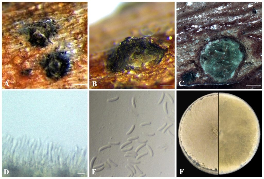
Pathogenic on branches of Prunus spp. Sexual morph: not observed. Asexual morph: Conidiomata 440-720 × 220-310 μm diameter, semi-immersed in host tissue, erumpent, discoid, solitary, circular to ovoid, scattered, multiloculate, with long ostiolar neck. Ostioles 125-160 μm diameter, at the same level as the disc. Conidiophores unbranched, reduced to conidiogenous cells. Conidiogenous cells 7.5–10 μm (n = 30), blastic, phialidic, originate from inner layer of pycnidial wall, hyaline, smooth-walled. Conidia (4.8–)5.5–7.2(–7.4) × 1.2–1.5(–1.7) μm (x̄ = 6.4 × 1.3 μm, n = 50), hyaline, allantoid, somewhat elongate, aseptate.
Material examined: CANADA, Ontario, Cedar Springs, N42°16’24.3”, W82°02’08.8”, isolated from main stem of Prunus armeniaca, 2018, O. Ellouz, K. Schneider, FDS-439; Lincoln, Jordan Farm (AAFC), 43°10’32.4″N 79°21’25.5″W, isolated from single conidia of Prunus persica var. nucipersica, Nov. 2019, E. Ilyukhin, O. Ellouz, FDS-564 (holotype); Lincoln, Jordan Farm (AAFC), 43°10’27.5″N 79°21’26.0″W, isolated from main stem of Prunus persica, Apr. 2021, O. Ellouz, K. Schneider, FDS-623.
Distribution: Canada.
Sequence data: ITS: OL640182, OL640183, OL640181 LSU: OL640184, OL640185, OL640123 ACT: OL631586, OL631587, OL631588 TEF -1a: OL631589, OL631590, OL631591
Notes: based on morphological characteristics and phylogenetic analysis, Cytospora paraplurivora is close to C. plurivora isolated from many different hosts in California (USA). This species is found to be associated with cancer disease of fruit (including Prunus spp.) and nut trees. Both species have common conidiomata and culture characteristics but conidia of C. paraplurivora are notably longer than those of C. plurivora (6.4 × 1.3 μm versus 4.1 × 1.0 μm) (Lawrence et al. 2018).

Fig. x. Cytospora paraplurivora on P. persica var. nucipersica (FDS-564). A. Habit of conidiomata on branches B. longitudinal section through conidiomata C. transverse section of conidiomata D. conidiogenous cells E. conidia F. seven-day-old culture on PDA. Scale bars: A= 200 μm B-C = 100 μm, D-E = 5 μm.
