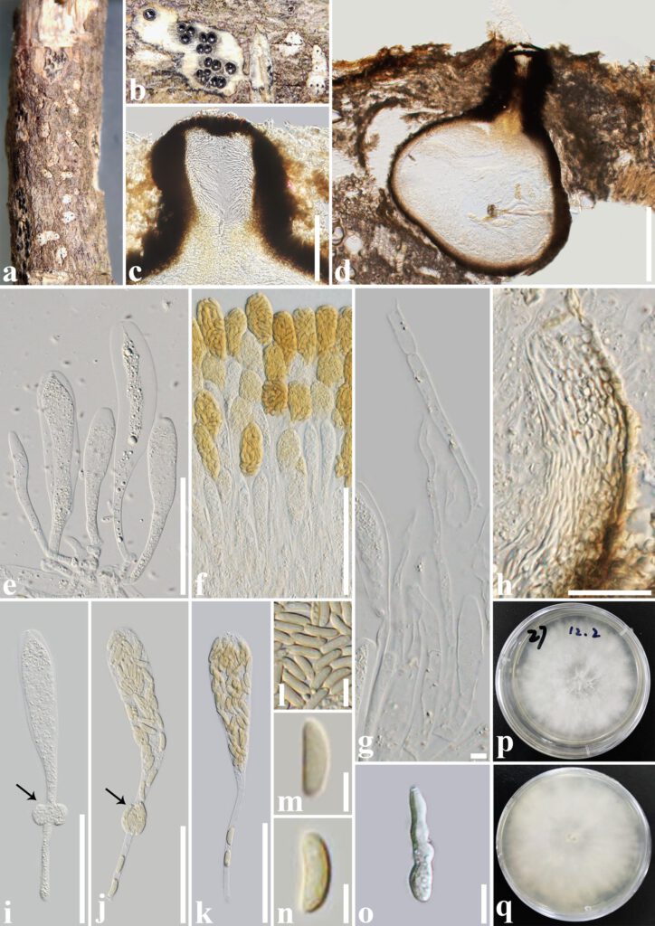
Allocryptovalsa aquilariae T.Y. Du & Tibpromma sp. nov.
MycoBank number: MB 846167; Facesoffungi number: FoF 12954; Fig. 80
Etymology – Named after the host genus, Aquilaria from which it was collected.
Holotype – HKAS 124187
Saprobic on dead twigs of Aquilaria sinensis (Lour.) Spreng. Sexual morph: Ascostromata gregarious with small black dots, immersed, the surrounding white host tissue without bark, 1–12-loculate. Ascomata (excluding the neck) 320–550 µm high × 250–650 µm diam. (x̄ = 380 × 410 μm, n = 10), perithecial, solitary to gregarious, immersed in substrate, globose to ampuliform, dark brown to black, not wrapped in white entostroma, the surrounding tissue is black after sectioning, ostiolate, papillate. Ostiolar canal 150–330 µm high × 85–150 µm diam. (x̄ = 203 × 112 μm, n = 10), central, not protrude or protrude slightly to outside from the substrate, cylindrical or irregular, straight, dark brown to black, periphysate. Peridium 35–95 μm wide, composed of several layers of thick-walled, hyaline to brown cells of textura angularis to textura prismatica, which are not fully fused with the host tissue. Hamathecium 2–9 μm wide, hyaline, with granulate, filamentous, unbranched, septate paraphyses, slightly constricted at the septa. Asci 130–190 × 10–20 µm (x̄ = 167.8 × 15.4 μm, n = 30), spore-bearing part length 65–100 μm (x̄ = 87 μm, n = 30), unitunicate, thin-walled, polysporous, clavate, J- apical ring, with 64–102 μm, apically rounded, narrowing towards lower region, with long and fragile pedicels, and some pedicels with subglobose or irregular structure. Ascospores (8–)9.5–11.5 × 2–3.5 µm (x̄ = 10.2 × 2.8 μm, n = 30), crowded, oblong to allantoid, pale yellowish at maturity, aseptate, slightly curved, smooth-walled. Asexual morph: Undetermined.
Culture characteristics – Colonies on PDA reaching 6 cm diam., after seven days at 28 ℃, flattened, filiform margin, with white aerial mycelia, flossy, velvety, reverse white, smooth.
Material examined – China, Yunnan Province, Xishuangbanna, dead twigs of Aquilaria sinensis, 13 September 2021, T.Y. Du, YNA27 (HKAS 124187, holotype), ex-type culture, KUNCC 22-10819, KUNCC 22-12389.
GenBank accession numbers – KUNCC 22-10819 – ITS: OP454035, β–tubulin: OP572197; KUNCC 22-12389 – ITS: OP456373, β–tubulin: OP572198.
Notes – In the phylogenetic tree, our isolates formed an inconspicuous branch with no support similar to Allocryptovalsa cryptovalsoidea, A. elaeidis, A. polyspora and A. truncata. However, the morphology of our isolates differs from the ones related to A. aquilariae (Fig. 1). Allocryptovalsa cryptovalsoidea has ostioles often perforated, emerging through the bark, while our species do not protrude or protrude slightly to the outside from the substrate, mostly immersed (Trouillas et al. 2011). The ascomata of our collection are 1–12-loculate, wrapped in white powder, which differs from the ascomata of A. elaeidis which have 1–2-loculate ascomata, delimited by a black zone in host tissues (Konta et al. 2020). Also, our species differs from A. polyspora by having 1–12-loculate, larger ascomata (320–550 × 250–650 µm vs. 80–425 × 100–400 µm), whereas the ascomata of A. polyspora are 1–3-loculate (Senwanna et al. 2017). Allocryptovalsa truncata has superficial ascostromata and individual ascomata, which differ from our collection by having 1–12-loculate, immersed ascostromata (Hyde et al. 2020). In addition, a PHI test, conducted based on the combined ITS and β-tubulin dataset showed that our Allocryptovalsa aquilariae isolates form a separate branch with Φw = 0.2266 (Φw > 0.05). Based on the multi-gene phylogenetic tree, PHI test results, and its unique morphological characteristics, Allocryptovalsa aquilariae is identified as a new species.

Fig. 1 — Allocryptovalsa aquilariae (HKAS 124187, holotype). a, b Appearance of ascostromata on the host. c Ostiolar periphysate. d Section through an ascoma. e, f, i-k Asci. g Paraphyses. h Peridium. l-n Ascospores. o A germinated ascospore. p, q Culture characteristic on PDA after seven days (p = colony in front, q = colony in reverse). Scale bars: c, f = 100 µm, d = 200 µm, e, h, i-k = 50 µm, g, m, n = 5 µm, l, o = 10 µm.
