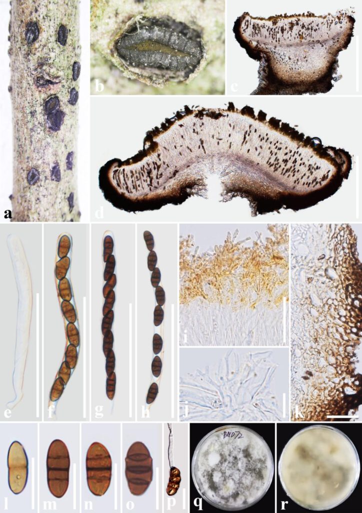
Rhytidhysteron bannaense T.Y. Du and Tibpromma sp. nov. (Figure 1)
MycoBank number: MB; Index Fungorum number: IF; Facesoffungi number: FoF 12957;
Etymology: Named after the region, where this sample was collected, Xishuangbanna.
Holotype: HKAS 122695
Saprobic on decaying wood of Buddleja officinalis (Loganiaceae). Sexual morph: Ascomata 1000–1500 μm long × 550–1100 μm wide × 350–850 μm high (x̅ = 1350 × 750 × 670 μm, n = 5), hysterothecial, solitary to aggregated, semi-immersed to superficial, black, apothecioid, navicular, rough, perpendicularly striate, elongate and depressed, compressed at apex, opening by a longitudinal slit, green at the center. Exciple 40–150 μm wide, composed of dark brown, thick-walled cells of textura angularis, outer layer brown to dark brown, inner layer pale brown to hyaline. Hamathecium comprising 1–2 μm wide, dense, hyaline, septate, branched, cellular pseudoparaphyses, forming an orange epithecium above asci when mounted in water, becoming purple epithecium above the asci when mounted in 10% KOH and turns hyaline after 5 seconds. Asci 140–189(–199) × 12–15(–16) μm (x̅= 166 × 14 μm, n = 20), 8-spored, bitunicate, cylindrical, with short pedicel, rounded at the apex, with an ocular chamber, J–. Ascospores 22–27 × 10–12.8 μm (x̅= 25 × 11.5 μm, n = 30), uni-seriate, slightly overlapping, hyaline when immature, becoming brown to dark brown when mature, ellipsoidal, rounded to slightly pointed at both ends, 3-septate, smooth-walled, without guttules and the mucilaginous sheath. Asexual morph: Undetermined.
Culture characteristics: Ascospores germinating on PDA within 24 h and germ tubes produced from one or both ends. Colonies on PDA reached a 6 cm diameter after two weeks at 28°C. The colony was soft, circular, irregularly raised, with an undulated edge, white to gray on the forward and grayish-yellow in reverse.
Material examined: China, Yunnan Province, Xishuangbanna Prefecture, Jinghong City, Manlie Village, on decaying wood of Buddleja officinalis Maxim. (Loganiaceae), 12 September 2021, T.Y. Du, BND72 (holotype, HKAS 122695, ex-type living culture, KUMCC 21-0482, living culture, KUMCC 21-0483).
Notes: Rhytidhysteron bannaense was closely related to Rhytidhysteron thailandicum Thambug. & K.D. Hyde and formed a well-supported clade (100% ML/1.00 PP). However, R. bannaense is distinct from R. thailandicum in having asci 8-spored, and ascospores 3-septate, without guttules, while asci (3–)6–8-spored, ascospores (1–)3-septate, guttulate in R. thailandicum (Thambugala et al. 2016, de Silva et al. 2020, Hyde et al. 2020a). In addition, the asci and ascospores size of R. bannaense are larger than those of R. thailandicum (Asci: 166 × 14 μm vs 145 × 12.8 μm; ascospores: 25 × 11.5 μm vs 24.5 × 9.5 µm) (Thambugala et al. (2016). Moreover, according to the comparison results of different gene fragments, R. bannaense is different from R. thailandicum in ITS and TEF genes (≥1.50%) (Table 3). Therefore, in this study, R. bannaense is introduced as a new species. Further details are given in Table 4.
Table 3. Comparison of LSU, ITS, LSU, and TEF gene fragments of R. bannaense and four strains of R. thailandicum. The “—” represents the non-available of this gene, superscripted T indicates ex-type.
| Closest known species | Strain number | LSU gene | ITS gene | SSU gene | TEF gene | References |
| R. thailandicum | MFLUCC 14-0503T | 0.00% | 1.38% | 0.00% | 2.19% | Thambugala et al. (2016) |
| MFLUCC 12-0530 | 0.00% | 1.54% | 0.00% | — | Thambugala et al. (2016) | |
| MFLU 17-0788 | 0.00% | 1.08% | 0.00% | — | de Silva et al. (2020) | |
| MFLUCC 13-0051 | 0.00% | 1.38% | — | 2.19% | Hyde et al. (2020a) |

Figure 1. Rhytidhysteron bannaense (HKAS 122695, holotype). a, b Appearance of hysterothecia on the host. c, d Vertical section through hysterothecium. e-h Asci. i Epithecium mounted in water. j Pseudoparaphyses. k Exciple. l-o Ascospores. p A germinating ascospore. q, r Colony on PDA medium (after four weeks). Scale bars: c, d = 500 μm, e-h = 100 μm, i, j, l-p= 20 μm, k = 50 μm.
