Fusarium solani (Mart.) Sacc., Michelia 2 (no. 7): 296 (1881)
Index Fungorum number: IF190352; Facesoffungi Number: FoF 01873
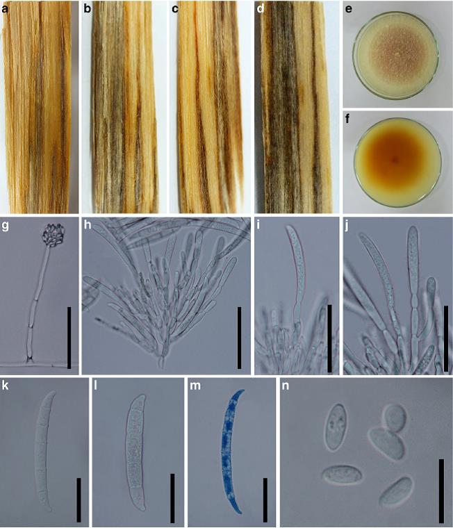
Associated with stem wilt disease of T. grandis. Sexual morph: Undetermined. Asexual morph: Colonies on PDA, Mycelium 2.4 4.2μm broad, branched, septate, slightly constricted at the septa, superficial, partly immersed, hyaline, verruculose. Conidiophores up to 190 μm long, 2–5.2 μm wide, micronematous, macronematous, densely branched, constricted at the septa, erect to slightly curved, up to 2–10- septate, Conidiogenous cell (7–)13–15(−17) × 3.5–5.1 μm, polyphialides, long, discrete. Macroconidia (40–)50– 52(−61) × 5–8 μm (x = 49×6μm, n = 30), hyaline, slightly curved, 4–6-septate, apical cells papillate, basal cells footshaped. Microconidia: (4–)6–7(−9) × 3.5–5 μm (x = 8.5 × 1.8μm, n = 30), subglobose, oval to truncate, aseptate, hyaline. Chlamydospores (8–)10–11(−10.6) × 7.5–11μm, aerial mycelia or embedded in the agar.
Culture characteristics: Pure culture was isolated by subbing hyphal tips growing from surface sterilized diseased material. Colonies on PDA reaching 59–66mmdiam. after 7 days in the dark at 25 °C, (x = 62, n =5), circular, edge entire, flat or effuse, aerial, medium sparse, light yellow (3A5) from above, light yellow (3A4) from below, reaching the edge of the Petridish after 10 days.
Habitat: Known to inhabit multiple plant families. Some strains may cause infections in humans (Farr et al. 2016) and associated with stem wilt disease of T. grandis (Doilom et al. 2017).
Known distribution: Cosmopolitan (Farr et al. 2016) and Thailand (Doilom et al. 2017).
Material examined: THAILAND, Chiang Rai Province, Muang District, Mae Fah Luang University campus grounds, associated with wilt disease of T. grandis stem, 19 November 2012, M. Doilom & J. Roux, living culture MFLUCC 12– 0770, MKT 088/1, GenBank Accession No: TEF1: KU315471; Chiang Rai Province, Muang District, associated
with wilt disease of T. grandis stem, 25 January 2013, M. Doilom, living culture MFLUCC 13–0329, MKT 101/1, ICMP 21172, GenBank Accession No: TEF1: KU315472; ibid. with MFLUCC 13–0329, living culture MFLUCC 13– 0338, MKT 105, GenBank Accession No: TEF1: KU315473.
Notes: The collections in this study are considered to be Fusarium solani based on TEE1 sequence data. Their morphology is similar to F. solani (Mart.) Sacc. (1881). Microconidia in this study are ((40–) 50–52 (−61) × 5–8 versus 40–60 × 7–8 μm) as reported in Sacc. (1881). The collections in this study were associated with wilt disease symptoms. Fusarium solani has previously been reported on teak in Tanzania (Ebbels and Allen 1979) and is illustrated here as it is the first report on T. grandis in Thailand, and to facilitate identification of this species on teak in future.
FIG: Fusarium solani (MFLU 15–3561). a–d Discolouration of stems. e, f Colony on PDA after 7 days (e = above view, f = below view). g Conidia attached to conidiophores. h–j Phialides. k–m Macroconidia. n Microconidia. Note: m Stained in lactophenol cotton blue. Scale bars: g–j = 100μm, k–m = 20μm, n = 10μm