Tubeufia tectonae Doilom & K.D. Hyde, sp. nov. Index Fungorum number: IF551973
Etymology: Name refers to the host genus Tectona on which the fungus was collected.
Holotype: MFLU 15–3423
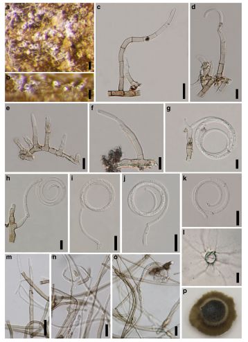
Saprobic on decaying inner-surface of bark of T. grandis. Sexual morph: Undetermined. Asexual morph: Colonies on natural substrate, superficial, gregarious, scattered, coiled, subhyaline to white. Conidiophores up to 125μm long, 3–6μm wide in broadest part, micronematous, slightly flexuous, septate, unbranched, filamentous, arising from hyphae or creeping hyphae, arising laterally from hyphae, simple, hyaline, pale brown at the base, gradually paler upwards, sometimes swollen at the base, indeterminate. Conidiogenous cells 7–15 long × 3.5–5μm wide in the broadest part, monoblastic or polyblastic, integrated, terminal. Conidia 32–55μm diam., coiled into 2–3 1/2 spirals, loose to tight, mostly loose, filaments 2–5μm wide, multi-septate, tapering towards rounded apex, acrogenous, helicosporous, hyaline to subhyaline, septate, conico-truncate at the base, expanded and uncoiled or loosely coiled (hygroscopic), inconspicuously constricted at the septa.
Culture characteristics: Conidia germinating on PDA within 24 h. Germ tubes produced around conidia. Colonies on PDA reaching 0.9–12 and 18–22 mm diam. after 7 and 14 days respectively in the dark at 25 °C (x = 10.7 and 20 mm after 7 and 14 days respectively, n = 5), edge entire to undulate, flat or effuse, sparse, slightly convex, with papillate surface at the old mycelium plugs, greyish brown (5D3) from both above and below. Mycelium 2–4.5 μm wide, partly superficial, partly immersed, pale brown to brown, septate, branched. Conidiophores up to 115 μm long, 2.5–6 μm wide, micronematous, slightly flexuous, septate, unbranched, filamentous, simple, hyaline, pale brown at the base, gradually paler upwards, sometimes swollen at the base, indeterminate. Conidiogenous cells 6–11 long × 3–4 μm wide in the broadest part, monoblastic or polyblastic, integrated, terminal or lateral. Conidia coiled into 1–2 1/2 spirals, loose to tight, mostly loose, filaments 2–5 μm wide, septate, multi-septate, tapering towards rounded apex, acrogenous, helicospore, hyaline to subhyaline, inconspicuously constricted at the septa, expanded and uncoiled or loosely coiled (hygroscopic). Chlamydospores developing in culture, catenate, intercalary or terminal, initially hyaline, becoming brown to dark brown at maturity, subglobose to ellipsoidal, thick-walled and smooth walled.
Habitat: Known to inhabit decaying inner-surface of bark of T. grandis (current study).
Known distribution: Thailand (current study).
Material examined: THAILAND, Chiang Rai Province, Muang District, Ban Doo Subdistrict, on decaying inner surface of bark of T. grandis, 15 July 2012, M. Doilom, (MFLU 15–3423, holotype), ex-type living culture MFLUCC 12–0392, MKT 065, ICMP 21162, GenBank Accession No: ITS: KU144923, LSU: KU764706, SSU: KU712460, TEF1: KU872763.
Notes: Based on phylogenetic analyses, Tu. tectonae in this study grouped within Tubeufia species. Tubeufia tectonae forms an asexual morph on the natural substrate, which is morphologically similar to Helicomyces, therefore, it was compared with known Helicomyces species. Tubeufia species have also been reported to produce helicomyces-like asexual morphs (Barr 1980; Samuels et al. 1979; Zhao et al. 2007), e.g. Tu. cylindrothecia has been identified as sexual morph of Helicomyces roseus (Barr 1980). Tubeufia tectonae is morphologically quite similar to H. colligatus, H. hyderbadensis and H. roseus, but its conidiophores are longer. Its conidia diameter is also wider than H. hyderbadensis. Colonies on natural substrata of Tu. tectonae are subhyaline to white but pale rose in H. colligatus, white to deep ash in H. hyderbadensis and white to pinkish in H. roseus. Tubeufia tectonae is introduced here as a novel species based on its morphological and phylogenetic differences from known Tubeufia and Helicomyces species
FIG Tubeufia tectonae (MFLU 15–3423, holotype). a, b Colonies on dead inner-surface of bark (a = top view, b = side view). c, e Conidiophores. d, f Developing conidia on conidiophores. g, h Conidiophores with conidia. i–k Conidia. l Germinated spore. m Developing conidium on conidiophore. n, o Conidiophores with conidia. m–o Morphology on MEA. p Close-up colony on PDA after 7 days. Scale bars a, b = 50 μm, c, i–k, m, n = 20 μm, d–h, o = 10 μm, l = 30 μm