Torula phytolaccae Y.X. Li, C.F. Liao & Doilom, sp. nov. (FIGURE 2)
MycoBank number: MB 559683; Index Fungorum number: IF 559683; Facesoffungi number: FoF 11438;
Etymology:—Name refers to the host genus Phytolacca on which the fungus was collected.
Holotype:—ZHKU 22-0056
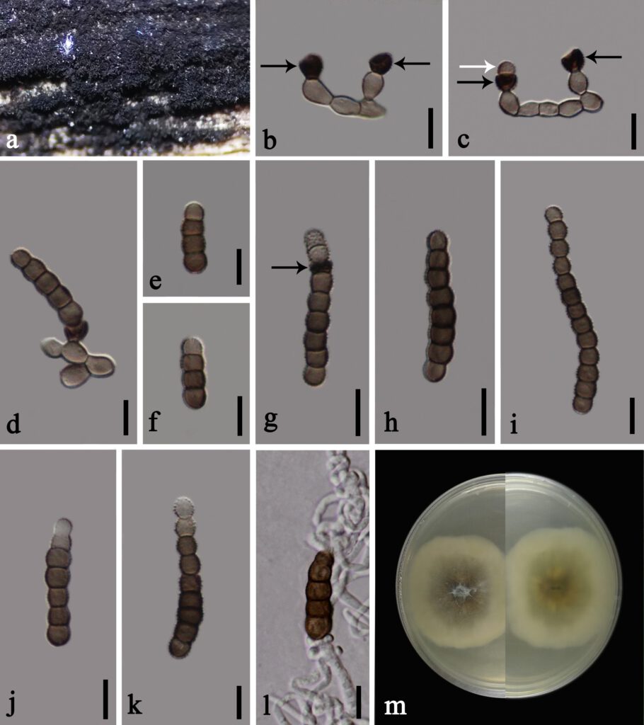
Saprobic on dead stems of Phytolacca acinosa. Sexual morph: undetermined. Asexual morph: Colonies effuse, dense, velvety, black on host. Mycelium 2–3 μm thick, immersed to superficial on the substrate, septate, branched, smooth to minutely verruculose, brown. Conidiophores 10–24 μm long × 3–7 μm wide ( = 18.5 × 5 μm, n = 15), semi-macronematous, mononematous, simple, flexuous, unbranched, verruculose, thick-walled, subcylindrical, consisting of 1–3 cells or reduced to conidiogenous cells, pale brown, arising from lateral and terminal on a hypha. Conidiogenous cells 4–6.7 μm long × 5–7.7 μm diam. ( = 5.3 × 6.5 μm, n = 20), solitary, monoblastic, lateral to terminal, dark brown to black, verruculose, thick-walled, cupulate. Conidia (9–)20–34(−63) × 5–9 ( = 27 × 6 μm, n = 40), phragmosporous, solitary to catenate, acrogenous, straight or slightly curved, pale brown to dark brown, verruculose, 2–12-septate, predominantly 3–5-septate, rounded at the both ends, composed of subglobose cells, constricted at the septa, occasionally subhyaline to paler brown at the apex, mostly subcylindrical, thick-walled.
Cultural characteristics:—Colonies on PDA reaching 28–30 mm diam. after 14 d at 25 ℃, medium dense, mycelium partly immersed to superficial, flat or effuse, edge entire, cottony, hairy at the centre, initially floccose at the centre with white and brown aerial mycelia, greyish green circle at the edge with pale creamy outward, becoming a purple-red circle, whitish at the margin from above; greenish grey the centre with pale creamy outward from below, without pigment produced in PDA. No asexual morph produced on PDA at 25 ℃ within 2 months.
Material examined:—CHINA, Yunnan Province, Kunming City, on dead stems of Phytolacca acinosa Roxb. (Phytolaccaceae), 28 June 2019, C.F. Liao, (ZHKU 22-0056, holotype); ex-type living culture ZHKUCC 22-0107, ibid., living culture ZHKUCC 22-0108.
Notes:—Morphologically, T. phytolaccae appears to have monoblastic conidiogenous cells, phragmosporous, and catenate conidia which are similar to T. thailandica. In addition, T. phytolaccae resembles T. hollandica and T. pluriseptata in having phragmosporous conidia composed of subglobose cells. However, T. phytolaccae differs from T. thailandica in having wider conidiophores (3–7 μm vs. 1.4–4.2 μm), the number of septa (2–12-septate vs. 2–8-septate), and longer and wider conidia ((9–)20–34(−63) × 5–9 μm vs. 14–23(–52.5) × 4.5–6.6 μm) (Hongsanan et al. 2020). Torula phytolaccae has cupulate conidiogenous cells and conidia are composed of subglobose cells, while T. thailandica has ellipsoidal conidiogenous cells and conidia are mostly composed of moniliform cells. Torula phytolaccae is distinct from T. hollandica in having more septa (3–12-septate vs. 2–4-septate). Torula phytolaccae has cupulate conidiogenous cells, while T. hollandica has doliiform conidiogenous cells. Torula phytolaccae differs from T. pluriseptata in having longer and wider conidiophores (10–24 μm long × 3–7 μm wide, consisting of 1–3 cells vs. (2.8–)3–4.3 μm long × 2.5–3 μm wide, consisting of 1–2 cells), the number of septa (3–12-septate, vs. 3–10-septate) (Li et al. 2017).
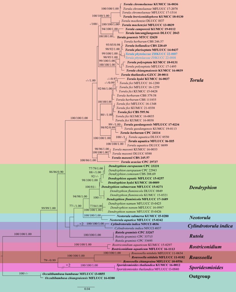
Phylogenetically, T. phytolaccae groups closed to T. herbarum (CBS 246.57: non-type), T. hollandica (CBS 220.69: ex-type), T. pluriseptata (MFLUCC 14-0437: ex-type), but forms a distinct linage with 98 % ML, 92 % MP, 1.00 PP support values (FIGURE 1). Considering these comparative details we introduce T. phytolaccae as a new species.

FIGURE 1. Maximum likelihood phylogenetic tree inferred from combined dataset of LSU, SSU, ITS, TEF1-α and RPB2 sequences of 67 taxa. Bootstrap support values for maximum likelihood (ML) and maximum parsimony (MP) equal to or greater than 75 % and Bayesian posterior probabilities (BYPP) equal to or greater than 0.95 are given above each branch in that order. The tree is rooted to Occultibambusa bambusae (MFLUCC 13-0855) and O. chiangraiensis (MFLUCC 16-0380). New isolates are shown in blue font. Ex-type strains are in bold.

FIGURE 2. Torula phytolaccae (ZHKU 22-0056, holotype). a Appearance of colonies on dead Phytolacca acinosa stem. b, c Conidiophores developed laterally and terminally on a hypha with dark conidiogenous cells (black arrows) and developed immature conidium (white arrow). d Conidium attached to conidiogenous cell. e–k Conidia. g Conidium with conidiogenous cells (black arrow) and attached immature conidium at the apex. l Germinated conidium. m Colony on PDA after 14 days (left:front, right:below). Scale bars: b–l = 10 μm.
