Tanmaurkiella Santam. gen. nov.
MycoBank number: MB 840611; Index Fungorum number: IF 840611; Facesoffungi number: FoF;
Diagnosis: Receptacle and primary appendage forming a common main axis without any evident transition between the receptacle and the primary appendage (i.e., the primary septum is not noticeable). Perithecium lateral, borne on a lower axial cell. Perithecial outer wall consisting of four vertical rows with four unequal cells each.
Etymology: Based on “Tanmaurk”, a name which appears in runic scripture on the larger of the two Jelling stones, which are strongly identified with the creation of Denmark as a national state and the origin for the actual name of Denmark.
Type species: Tanmaurkiella pselaphi Santam.
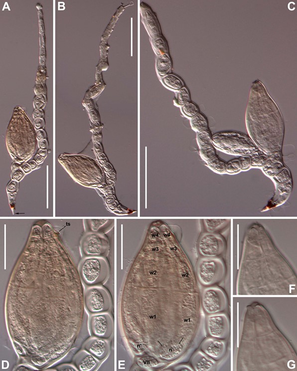
Description: Receptacle and primary appendage forming a common, continuous, long axis. The exact position of the primary septum could not be determined, therefore cell III cannot be properly described. Perithecium lateral, usually arising from the second or third axial cell, and separated from them by a vertical septum; if any additional perithecium is formed it arises from cells above. Perithecial basal cells (m, n, n’) and secondary perithecial stalk cell (VII) with well-distinguished walls at maturity. Perithecial outer wall consisting of four vertical rows, each with four cells of unequal height, the cells of each row progressively shorter towards the perithecial tip. Perithecial apex ornamented with lobes developed by tier w4 cells (Fig. 63F–G). Antheridia unknown.
Remarks: This genus seems related with Bordea and Cryptandromyces according to certain morphological and ecological affinities. These genera, like Tanmaurkiella, infect Pselaphinae beetles (Col. Staphylinidae). Bordea and Cryptandromyces may be separated from Tanmaurkiella by one to several characteristics. Species of Bordea show a different appendage arrangement, with three superposed cells, including a distal antheridium; nevertheless, the perithecial apex of species in Tanmaurkiella remind those of Bordea by the presence of a crown-like structure. The genus Cryptandromyces includes species with three- celled receptacles and with perithecia where the outer wall shows five cells of subequal height in each vertical row of cells. Also, the new genus might be morphologically compared with some species of Siemaszkoa, like S. ptenidii, by the organization of the receptacle and appendage forming a continuous axis, with clearly lateral perithecia; nevertheless, this may be considered a homoplasy because remaining characteristics are very different, especially those dealing with the perithecium.

Fig. 63. Tanmaurkiella huggertii Santam. gen. et sp. nov. A–B. Mature thalli. In A, the pointed hyaline beak at the base of the foot is labelled (arrow). C. Mature thallus with a second young perithecium. D–E. Perithecium in detail, at two focus levels, near (D) and far (E), to show perithecial wall cells (wn) developed from cell “n”, cells n’, VII and trichogyne scar (ts). F–G. Perithecial tip in detail at near (F) and far (G) focus to show the four preapical protuberances. Scale bars: A–C = 50 µm; D–G = 20 µm. Photographs from slide ZMUC C-F-122720 (holotype).
Species
