Tahromyces munnarensis Hanafy, Vikram B. Lanjekar, Prashant K. Dhakephalkar, T.M. Callaghan, Dagar, G.W. Griff., Elshahed & N.H. Youssef, sp. nov.
MycoBank number: MB 830866; Index Fungorum number: IF 830866; Facesoffungi number: FoF;
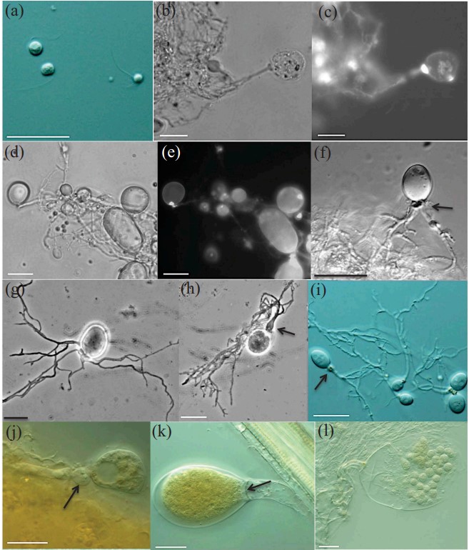
Typification: The holotype (FIG. 9h) was derived from the following: INDIA. KERALA: Town of Munnar, 10.219°N, 77.106°E, ~2100 m asl, 3-d-old culture of isolate TDFKJa193, originally isolated from freshly deposited feces of a Nilgiri tahr (Nilgiritragus hylocrius), Sumit Dagar. Ex-type strain: TDFKJa193. GenBank: MK775321 (ITS1); MK775310 (D1–D2 28S rDNA).
Etymology: The species epithet “munnarensis” refers to the town that the type species was isolated from. Obligate anaerobic fungus that produces globose monoflagellate zoospores. Biflagellate and triflagellate spores are rarely identified. Both endogenous and exogenous sporangia were observed. The sporangia vary in size between 12 and 100 μm in length and between 10 and 70 μm in width and display a wide range of morphologies such as globose, ovoid, and obovoid. Sporangiophores are short (12 to 20 µm) with frequent subsporangial swellings. Sporangial necks (1 to 8 μm W and 2 to 10 μm L) are frequently constricted. Septa often form at the base of mature exogenous sporangia.
Zoospore liberation happens after irregular dissolution of the sporangial wall. Produces colonies that are small (1 mm), white in color with a compact and fluffy center, surrounded by dotted circles of fungal thalli. In liquid media, it produces numerous fungal thalli attaching to the glass bottles on initial days of growth and later developing into thin, mat-like structures. The clade is defined by the sequence MK775310 (D1–D2 28S rDNA).
Additional specimens examined: INDIA. KERALA: Town of Munnar, 10.219°N, 77.106°E, ~2100 m asl, 3-d-old culture of isolates TDFKJa1924, TDFKJa1926, and TDFKJa1927, originally isolated from freshly deposited feces of a Nilgiri tahr (Nilgiritragus hylocrius), Sumit Dagar. GenBank (D1–D2 28S rDNA): TDFKJa1924(MK775312.1),TDFKJa1926 (MK775311.1), and TDFKJa1927 (MK775313.1).

Figure 9. Microscopic features of Tahromyces munnarensis (clade 7, Nilgiri tahr) strain TDFKJa193 (W-KE). Differential interference contrast (a, f, i–l), phase-contrast (b, d, g, h), and fluorescence (c, e) micrographs. a. Monoflagellate and triflagellate zoospores. b–e. Monocentric thalli; nuclei were observed in sporangia, not in rhizoids or sporangiophores. f–g. Endogenous sporangia: f. Ovoid endogenous sporangium with two rhizoidal systems; note the subsporangial swelling (arrow). g. Endogenous sporangium with branched rhizoids. h–l. Exogenous sporangia: h. Globose sporangium with short swollen sporangiophore (arrow). i–j. Exogenous sporangia with subsporangial swellings and constricted necks (arrows). k. Ovoid sporangium with septum at the sporangial base (arrow). l. Zoospore release through dissolution of a wide apical pore. Bars = 20 μm.
