Stilbochaeta malaysiana (Kuthub.) Réblová & Hern.-Restr., comb. nov.
MycoBank number: MB 842216; Index Fungorum number: IF 842216; Facesoffungi number: FoF; (Figures 34 and 38).
Basionym. Dictyochaeta malaysiana Kuthub., Trans. Br. mycol. Soc. 89: 356. 1987.
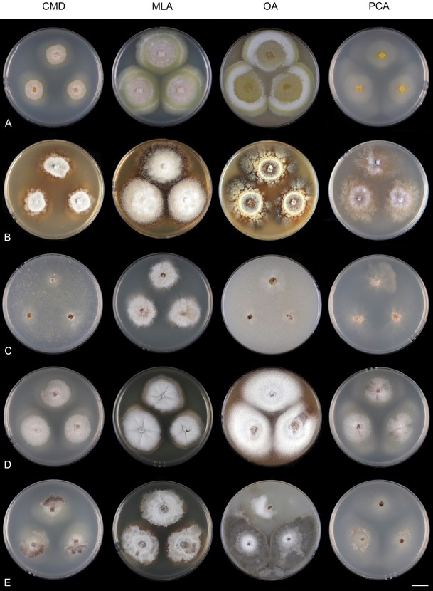
Culture characteristics: On CMD: colonies 24–25 mm diam, circular, raised, margin undulate, lanose, cobwebby at the margin, grey-beige centrally, beige toward the margin, ochre pigment diffusing into agar, reverse dark cinnamon. On MLA: colonies 62–63 mm diam, circular, raised, flat margin, margin fimbriate, lanose, zonate, finely furrowed, whitish-beige becoming olivaceous ochre-beige toward the periphery, dark cinnamon at the margin, caramel pigment diffusing into the agar, reverse dark cinnamon. On OA: colonies 61–62 mm diam, circular, raised, margin lobate, lanose, floccose, furrowed, zonate with a central cobwebby ring surrounded by small lanose areas of radially spreading mycelium, ochre-grey centrally, pale ochre-beige to olivaceous cinnamon toward the periphery, aerial hyphae with numerous pale ochre exudates, dark cinnamon pigment diffusing into the agar, reverse dark amber to brown. On PCA: colonies 32–38 mm diam, flat, circular, margin undulate, sparsely lanose to cobwebby, ochre-beige, pale cinnamon pigment diffusing into the agar, reverse cinnamon. Sporulation was moderate on CMD and PCA, absent on MLA and OA.
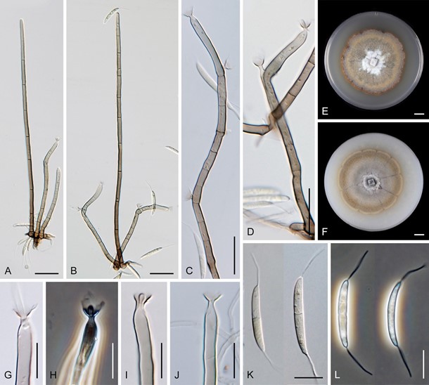
Colonies on CMA with U. dioica stems effuse, hairy, mycelium composed of branched, septate, hyaline to subhyaline hyphae 1.5–3 µm diam. Anamorph. Setae 230–281 µm long, 4–5.5 µm wide near the swollen base, single or arise in groups of two from knots of dark brown hyphal cells, erect, straight or flexuous, septate, smooth, dark brown and thick- walled, paler and thinner-walled toward the apex, unbranched, apex pale brown, sterile. Conidiophores 78–113 µm long, 4–5.5 µm wide near the base, macronematous, single or arise in fascicles of 2–5 from knots of dark brown hyphal cells around the base of the setae, erect, straight or bent, septate, smooth, medium to pale brown, paler toward the tip. Coni- diogenous cells 17–21(–37) × 3.5–4.5(–5) µm tapering to 1.5–2 µm below the collarette, inte- grated, terminal, mono- or polyphialidic with 1–3 lateral openings while internally septa can be formed, extending percurrently and sympodially, subcylindrical, pale brown; col- larettes 4.5–7 µm wide, 3.5–4.5 µm deep, funnel-shaped to slightly campanulate, tubular at the base, pale brown. Conidia 21–26 × 2.5–3.5 µm (mean ± SD = 23.6 ± 1.3 × 3.0 ± 0.2 µm), falcate, with an inconspicuous basal scar, 1-septate, hyaline, with a straight or gently curved setula at each end, 12–18 µm long, positioned terminally at the apex and slightly subtermi- nally at the base, conidia accumulate in slimy whitish fascicles. Teleomorph. Unknown.
Specimen examined: MALAYSIA, Selangor, Kepong Forest Reserve, on decaying leaves, October 1986, A.J. Kuthubutheen KUM 534 (holotype IMI 312436, ex-type strain IMI 312436).
Habitat and geographical distribution: Saprobe on decaying leaves known from Southeast Asia, Malaysia [19]. According to GlobalFungi, the identical sequences were found in 13 soil samples collected in forests of the Caribbean (12 samples, Puerto Rico, Trinidad and Tobago [119,185] and South Asia (one sample, India) [117]. The locations have tropical climate (MAT avg. 22 ◦C, MAP 3124 mm).
Notes: Kuthubutheen [19] listed two specimens with different localities in the pro- tologue, however, only one number to indicate the holotype IMI 312436. By comparing the protologue with the type material deposited in the Kew herbarium, it was found that the locality of a specimen listed second in order is the place of collection of the holo- type. Kuthubutheen [19] did not indicate an ex-type strain of his new species. Strain of S. malaysiana, with identical designation IMI 312436 as the holotype and accompanying metadata, deposited by A.J. Kuthubutheen, was obtained from the CABI culture collection. We confirm its identity and connect the holotype with the ex-type strain in this study.

Figure 34. Colony morphology of Stilbochaeta spp. after 4 weeks. (A) S. aquatica CBS 114070 (B) S. malaysiana IMI 312436 ex-type (C) S. ramulosetula IMI 313452 ex-epitype (D) S. septata CBS 143386 ex-epitype (E) S. septata CBS 146716. Scale bar: (A–E) 1 cm.

Figure 38. Stilbochaeta malaysiana (IMI 312436 ex-type). (A,B) Setae and conidiophores (C,D) conidiophores with conidio- genous cells, in detail (E,F) colonies (G–J) tip of the conidiogenous cells with collarettes (K,L) conidia. Images: (A–D,G–L) on CMD after 4–6 weeks (E) on MLA after 4 weeks (F) on OA after 4 weeks. Scale bars: (A,B) 25 µm; (C,D) 20 µm; (E,F) 1 cm; (G–L) 10 µm.
